Download
1 / 17
170 likes | 179 Views
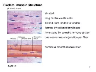
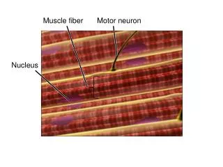
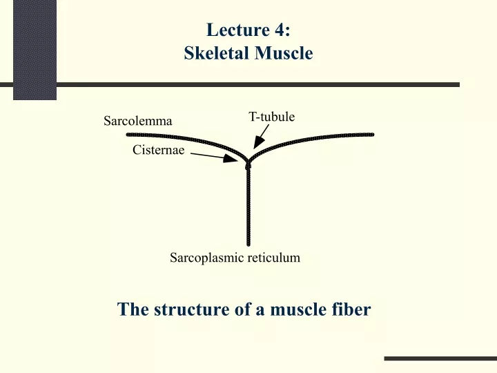
T-tubule. Sarcolemma. Cisternae. Sarcoplasmic reticulum. Lecture 4: Skeletal Muscle. The structure of a muscle fiber. Cross bridges. Z-line. Actin. Myosin. A-band. I-band. A-band. I-band. A-band. Structure of a Myofibril.

E N D
T-tubule Sarcolemma Cisternae Sarcoplasmic reticulum Lecture 4: Skeletal Muscle The structure of a muscle fiber
Cross bridges Z-line Actin Myosin A-band I-band A-band I-band A-band Structure of a Myofibril The lower figure shows the sequence of dark and light bands. The upper figure shows the typical configuration of actin and myosin molecules within a myofibril.
Tropomyosin Troponin Actin Structure of the Thin Filament (Actin) Note long tropomyosin molecules in parallel with the actin strands. Troponin attaches to tropomyosin at regular intervals.
Nerve action potential Synaptic vesicles ACh Presynaptic membrane AChesterase Motor end plate Neuromuscular Synapse A presynaptic nerve action potential induces movement of vesicles with acetylcholine (ACh) to the presynaptic membrane, their fusion, and release of ACh into the cleft. ACh diffuses to the postsynaptic muscle membrane, depolarizes it, and induces an action potential.
Neuromuscular Synapse • Neurotransmitter: acetylcholine (ACh) • Always excitatory • Obligatory • No multiple innervation • AChesterase quickly destroys ACh in the synaptic cleft
V Time Spontaneous MEPPs Nerve action potential Muscle AP Miniature excitatory postsynaptic potentials (end plate potentials, MEPPs) spontaneously occur on the postsynaptic muscle membrane. A presynaptic nerve action potential always reaches the depolarization threshold and induces a muscle action potential.
Sarcolemma ++ ++ Ca Ca Sarcoplasmic reticulum Direct Effects of the Muscle AP Muscle action potential travels along the sarcolemma, enters T-tubules, and leads to a release of Ca++ ions from the sarcoplasmic reticulum.
Cross bridge ++ Ca Actin Tropomyosin Troponin ATP Myosin Sliding Filament Theory Ca++ ions remove tropomyosin and free a site for myosin to bind to troponin (this process uses the energy from ATP). A ratchet motion occurs, moving the filaments with respect to each other.
Force Latent period Time 100 ms Muscle Twitch A typical twitch contraction of a muscle in response to a single stimulus.
Force Time Temporal Summation of Muscle Twitches Two action potentials come at a short interval and induce two twitch contractions. Their mechanical effects are superimposed, leading to a higher level of muscle force.
Force Smooth tetanus Time Action potentials Tetanus A sequence of action potentials may lead to a tetanus (a sustained contraction). At a high frequency of action potentials, individual contractions may fuse, leading to a smooth tetanus.
A Bit of Basic Mechanics • Stiffness: a property of a spring, a structure that deforms and accumulates potential energy under the influence of an external force F = −k*∆x • Damping (“viscosity”): a property of an object to generate force against a velocity vector F = −b*V • Inertia: a coefficient of proportionality between force and acceleration F = m*a
K1 B F K2 Force A Simple Hill-Type Muscle Model A simple mechanical model of a muscle. It contains a force generator (F), a viscous element (B), and two elastic elements: a parallel spring (K1) and a series spring (K2).
Force S1 S2 S3 Length Spring Properties of an Isolated Muscle Force-length curves measured in a muscle for different levels of external stimulation (S1, S2, and S3). The muscle behaves like a nonlinear spring. Changing the strength of the stimulation modifies the zero length of the spring.
Velocity Force 0 F 0 Force-Velocity Muscle Properties A typical force-velocity curve for a whole muscle. According to tradition, the Y-axis represents the velocity of muscle shortening. The muscle produces higher forces when it is lengthening (negative velocity) than when it is shortening (positive velocity). Compare this figure with the Hill equation.
Load Isometric Muscle characteristic Elastic Isotonic Length External Loads A muscle always works against a load. Three types of loads are illustrated: an isometric load prevents changes in “muscle plus tendon” length; an isotonic load does not change; and an elastic load acts like a spring. A typical muscle characteristic (the thin curve) is shown for comparison.
Regimes of Muscle Contraction • Concentric: A muscle develops force while shortening. • Eccentric: A muscle develops force while lengthening. External loads: • Isometric: The “muscle plus tendon” length does not change. • Isotonic: The apparent external load does not change. • Elastic: The load is a spring.