Download
1 / 20
460 likes | 1.08k Views
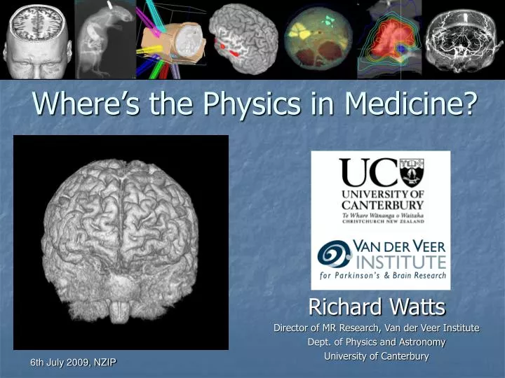
Where’s the Physics in Medicine?. Richard Watts Director of MR Research, Van der Veer Institute Dept. of Physics and Astronomy University of Canterbury. Therapy. Therapy. MRI. Nuclear Imaging. X-Ray. Diagnostic Imaging. X-ray source. Detector. Object.

E N D
Where’s the Physics in Medicine? Richard Watts Director of MR Research, Van der Veer Institute Dept. of Physics and Astronomy University of Canterbury
Therapy Therapy MRI Nuclear Imaging X-Ray Diagnostic Imaging
X-ray source Detector Object X-Ray ImagingContrast: Density, Atomic Number December 22nd, 1895 Wilhelm Roentgen “Builder survives nailgun accident” Phil and Anthony Butler, UC and MARS Bioimaging
Pneumoencephalography Pneumoencephalogram from Moore et al (1935) 'Encephalographic studies in mental disease' - American Journal of Psychiatry
Medipix – Spectroscopic X-ray Imaging (CERN)Separate Density and Atomic Number “Colour X-ray Imaging”
Computed Tomography (CT)3D X-Ray Imaging Prototype MARS Spectroscopic CT scanner CT Scanner rotating
Nuclear Imaging, Gamma Camera (1957)Contrast: Concentration of radiopharmaceutical
Radiopharmaceuticals for Nuclear Imaging EC = Electron capture, IT = Isometric transition
Single Photon Emission Computed Tomography, SPECT3D Nuclear Imaging Bushberg, Essential Physics of Medical Imaging
Positron Emission Tomography (PET) 18F-FDG, T1/2 ~ 2 hours
Hydrogen Nuclei are aligned by a big Magnetic Field Resonant frequency given by Larmor equation Nuclei absorb radiofrequency energy at that frequency and then re-emit energy Magnetic field gradient coils allow the field (and resonant frequency) to vary with location in x,y,z – Imaging Magnetic Resonance Imaging, (n)MRI “You know, what these people do is really clever. They put little spies into molecules and send radio signals to them, and then they have to radio back what they are seeing.” Niels Bohr
Structural Brain Imaging with MRI T1 T2 FLAIR T2 Apparent Diffusion Coefficient Fractional Anisotropy T2*
Functional MRI of Language – Passive Listening, Rhyming • Magnetic properties of oxyhemoglobin and deoxyhemoglobin • L. Pauling and C. Coryell, PNAS USA 22:210-216 (1936)
Radiation therapy Normal tissue complication probability Tumour control probability Cure Response Dose
Intensity-Modulated Radiotherapy (IMRT) High Dose Region Constrained inverse problem Tumour Patient Intensity Modulated Cross - section Beam Profiles Linear Accelerator (linac) Juergen Meyer, UC
Summary • Physics has made, and continues to make essential contributions to modern medicine • Imaging • X-ray imaging (Medipix/MARS) • Nuclear imaging • MRI (Functional imaging) • Radiation therapy • Accelerator physics • Treatment planning, Monte Carlo modeling • “Blue sky” research results in important unexpected technology (e.g. PET) • Medical physics • Excellent career prospects • Using physics to improve health