Download
1 / 24
310 likes | 477 Views
Gain comprehensive knowledge of liver anatomy, functions, diseases, and diagnosis related to jaundice. Explore differential diagnosis, lab estimations, and a structured approach to understanding this condition. Learn from the expertise of Dr. Ravi Vaswani, MD, a Professor at Yenepoya Medical College, Mangalore.

E N D
Jaundice - A Clinical Approach Dr. Ravi Vaswani MD Professor, Department of Medicine Yenepoya Medical College, Mangalore
Liver anatomy • Largest organ • 1-1.5 kg • 20% blood flow from hepatic artery - oxygen rich • 80% from portal - nutrient rich • Hepatocytes 66% • Kuppfer (RE), Ito (fat-storing), endothelial, bile ductular Ravi Vaswani MD
Blood flow patterns Ravi Vaswani MD
Anatomical lobular structure Ravi Vaswani MD
Functional acinar structure Ravi Vaswani MD
Flow patterns • Blood from portal triad flows towards central hepatic vein (across zones 1, 2, 3) • Bile - secreted by hepatocytes - flows in a counter current (zones 3, 2, 1) • Hepatic sinusoids lined by cells that have loose junctions allow plasma (not cellular elements) to flow into the subendothelial space of Disse Ravi Vaswani MD
Functions • Synthesis of essential serum proteins (albumin, carrier proteins, coagulation factors, hormonal/growth factors • Production of bile and its carriers (bile acids, cholesterol, lecithin, phospholipids) • Regulation of nutrients (glucose, glycogen, lipids, amino acids) • Metabolism, conjugation of lipophilic compounds (bilirubin, anions, cations, drugs) Ravi Vaswani MD
Liver function tests • Bilirubin, ser albumin and prothrombin time are the most commonly assessed LFT • Bilirubin is a measure of hepatic conjugation and excretion • Albumin & Prothrombin time are measures of protein synthesis Ravi Vaswani MD
Classification of liver diseases • Hepatocellular (functions are suboptimal) • Viral hepatitis, alcoholic liver disease • Obstructive (Cholestatic) (bile flow stopped) • Gallstones, malignancies, primary biliary cirrhosis, drug-induced liver disorder • Mixed • Cholestatic stage of viral hepatitis • Drug-induced liver disorder Ravi Vaswani MD
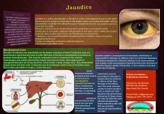
Introduction • Yellowish discoloration of tissues (sclera, skin and mucus membranes) from deposition of bilirubin • Occurs only if serum hyperbilirubinemia • Either liver disease or hemolytic disorder • Slight increases in serum bilirubin are best detected by examining the sclerae, which have a particular affinity for bilirubin due to their high elastin content (procollagen IV) Ravi Vaswani MD
Intro contd… • Scleral icterus indicates a serum bilirubin of at least 3.0 mg/dL • A second place to examine is underneath the tongue • As bilirubin levels rise, skin will eventually become yellow in light-skinned patients & green if the process is long-standing due to oxidation of bilirubin to biliverdin • Confirm by examining urine (dark yellow) Ravi Vaswani MD
Differential Diagnosis • Carotenoderma: yellow color imparted to skin due to carotene • Use of the drug quinacrine • Excessive exposure to phenols • In jaundice, yellow discoloration of skin is uniformly distributed over the body • In carotenoderma the pigment is concentrated on palms, soles, forehead, & nasolabial folds. • Carotenoderma can be distinguished from jaundice by the sparing of the sclerae. Ravi Vaswani MD
Lab Estimation • The terms direct- & indirect-reacting bilirubin are based on the original van den Bergh reaction • Bilirubin is exposed to diazotized sulfanilic acid, splitting into two stable dipyrrylmethene azopigments that absorb maximally at 540 nm, allowing for photometric analysis • Direct fraction reacts with diazotized sulfanilic acid in the absence of an accelerator substance such as alcohol & provides an approximate determination of conjugated bilirubin Ravi Vaswani MD
Total serum bilirubin is the amount that reacts after the addition of alcohol. • Indirect fraction is the difference between total and direct bilirubin; provides an estimate of unconjugated bilirubin in serum. • In van den Bergh method, normal serum bilirubin concentration is 17 mol/L (<1 mg/dL) • Up to 30%, or 5.1 mol/L (0.3 mg/dL), of the total may be direct-reacting (conjugated) bilirubin. • Total serum bilirubin concentrations are between 3.4 and 15.4 mol/L (0.2 and 0.9 mg/dL) Ravi Vaswani MD
Bilirubin in the Urine • Unconjugated bilirubin is bound to albumin; is not filtered by kidney, and is not found in urine • Conjugated bilirubin is filtered – glomerulus; majority is reabsorbed by proximal tubules; a small fraction is excreted in the urine • Bilirubin found in urine is conjugated bilirubin. • Bilirubinuria implies liver disease • A urine dipstick test (Ictotest) gives the same information as fractionation of the serum bilirubin Ravi Vaswani MD
The Approach • Hyperbilirubinemia may result from • Overproduction of bilirubin • Impaired uptake • Impaired conjugation • Impaired excretion • Regurgitation of unconjugated or conjugated bilirubin from damaged hepatocytes or bile ducts Ravi Vaswani MD
Unconjugated hyperbilirubinemia results from either overproduction, impairment of uptake, or conjugation of bilirubin • Conjugated hyperbilirubinemia is due to decreased excretion into the bile ductules or backward leakage of the pigment Ravi Vaswani MD
Clinical approach - Step 1 • Take detailed history; focus on drug intake • Past history of blood transfusion, jaundice • History of alcohol intake (80 gm for 10 years or 160 gm for 5 years) • Do a physical examination: look for hepatomegaly, splenomegaly, ascitis, lymph nodes, s/o chronic hepatocellular failure Ravi Vaswani MD
Step 2: Liver Function Tests (LFT) • Bilirubin (total and direct) • AST & ALT • Prothrombin time • Proteins • Alkaline phosphatase Ravi Vaswani MD
Step 3 • Is the hyperbilirubinemia predominantly conjugated or unconjugated in nature? Step 4 • Are other liver biochemical tests abnormal? Ravi Vaswani MD
Isolate direct hyperbilirubinemia • Consider • Dubin-Johnson syndrome • Rotor’s syndrome Ravi Vaswani MD
Isolated indirect hyperbilirubinemia • Consider • Gilbert syndrome • Crigler-Najjar syndrome • Hemolytic disorders • Drugs (Rifampicin, probenecid) Ravi Vaswani MD
Combined hyperbilirubinemia with other biochemical tests abnormal • AST & ALT out of proportion to Alk phos; consider hepatocellular pattern; viral markers, ceruloplasmin, tox profile • Alk phos out of proportion to AST/ALT consider cholestatic pattern; USG to rule out obstruction • CT abdomen if indicated • Consider liver biopsy Ravi Vaswani MD
Tips • Low albumin suggests chronic process such as cirrhosis or cancer • Normal albumin suggests more acute process such as viral hepatitis or choledocholithiasis • ↑ PT indicates either vitamin K deficiency due to prolonged jaundice and malabsorption of vitamin K or significant hepatocellular dysfunction • Failure of PT to correct with parenteral admin of vitamin K indicates severe hepatocellular injury Ravi Vaswani MD