Download
1 / 37

E N D
Jaundice Dr. Ahmed Kensarah
Introduction Surgical obstructive jaundice (jaundice due to intra- or extra-hepatic organic obstruction to biliary outflow) can present problems in diagnosis and management. This is so because, there is a hard core of jaundiced patients in whom it is very difficult to distinguish between organic obstruction and medical causes of jaundice, particularly intrahepatic cholestasis. Even serial liver function tests are often inconclusive in differentiating However, it is mandatory to determine pre-operatively the existence, the nature and the site of obstruction in the surgical cases because an ill chosen therapeutic approach can be dangerous.
Material and Methods Twenty-six consecutive cases of obstructive jaundice were diagnosed and treated in one full-time surgical unit over a period of 3 years from 1976 to 1979. Of these, 14 cases had malignancy and 12 cases belonged to the non-malignant group. A11 the patients were above 40 years of age and the male: female ratio was 1.9:1.The patients were subjected to a detailed clinical examination particularly with reference to the enlargement of liver, spleen and gall bladder. They also had urine examination, hemogram and, serum chemistry including liver function tests.
Material and Methods (cont.) Australia antigen examination was done in 15 cases. Citrate clearance test' was done in 12 patients. All the patients had plain X-ray of abdomen and upper GI Barium series. Oral cholecystography was done in 7 patients whose serum bilirubin was less than 3 mg%,-. Percutaneous transhepatic cholangiography (PTC) was done in 9 patients. Liver scan using 99mTc phytate was done in 13 cases. Selective hepatic angiography was done pre-operatively in 2 patients.
Material and Methods (cont.) The patients were prepared for surgery with injectable Vitamin K to correct the prothrombin time; they were given fresh blood transfusions if the prothrombin time did not improve. In order to avert possible post-operative renal failure, all patients were treated with correction of dehydration, intravenous Mannitol and intravenous Frusemide pre-operatively.
Material and Methods (cont.) PTCD-Percutaneous Transhepatic Cholangiography with drainage. The patients were treated with various surgical procedures as shown in [Table 1]. Some of the patients had more than one surgical procedures mentioned in the table. Curative surgery was attempted in benign conditions and in early malignancies. In the advanced malignancies; surgery was mainly palliative.Whenever bile could be obtained either during P.T.C. or during laparotomy (20 cases), it was subjected to bacteriological examination.
Material and Methods (cont.) Intra-operative cholangiography was done in 3 cases and it showed the sites of obstruction. Tube cholangiography was done post-operatively in 11 cases either through the cholecystostomy tube or through a splint kept in the biliary tree.
Results (cont.) The patient with hepatoma of the liver and one patient with carcinoma of the gall bladder infiltrating into the liver had hard enlarged liver. There were 6 cases who illustrated an exception to Courvoisiers law.Of these, 4 were patients with cholelithiasis and a palpable gall bladder; of these, 2 had an associated malignancy of the biliary tract. The remaining 2 exceptions were patients with malignant obstruction of the lower end of the common bile duct (CBD), in whom the gall bladder was not palpable; in one of them, this was due to an associated carcinoma of the right hepatic duct involving the cystic duct.
Results (cont.) Nineteen out of the 26 patients had serum albumin level of less than 3 gms per cent. The average total serum bilirubin was 10.4 mg%, the highest being 35.5 mg%; and the lowest being 1.6 mg%. The SGPT' was elevated (more than 40 Reitman and Frankel units/ml) in 11 patients; it was more than 1G0 Reitman and Frankel Units /ml in 10 patients. The alkaline phosphatase was elevated (more than 30 K.A. units) in 19 patients; it was normal in 7 patients. The prothrombin time was elevated (more than 16, seconds) in all patients. Citrate clearance was abnormal in all the patients.
Results (cont.) Plain X-ray abdomen showed enlarged liver shadows in 8 patients and radio opaque gall stones in 5 patients. Barium meal examination of the G.I. tract showed chronic gastritis with duodenal ulcer in 1 case of gall stones, indentation of duodenum by enlarged common bile duct in 3 patients, `inverted three' (8) appearance in periampullary malignancy (1 case), widening of duodenal C in 2 cases of carcinoma of head of pancreas and displacement of the stomach by enlarged liver in 1 case of hepatoma of the liver.Oral cholecystography showed filling defects suggestive of stones in 2 patients and failure of visualisation of the gall bladder in 4 patients; it was normal in 1 patient whose serum bilirubin was 1.6 mg%.
Results (cont.) PTC showed obstruction at the lower end of the CBD in 5 cases, 2 due to stones, 2 due to malignancy and one due to inflammatory stricture. PTC also helped to diagnose choledochal cyst in 2 cases (which showed dilated CBD) and it showed dilated intrahepatic ducts filled with stones in 2 cases.
Results (cont.) Selective hepatic angiography showed an avascular area in the patient with intrahepatic choledochal cyst and in the patient with hepatoma, it outlined the vascular tumor.Hepatic scanning showed mild to moderate hepatomegaly in 12 cases. Two patients showed cold areas in the liver and another 2 in the region of the gall bladder invaginating into the liver substance suggesting a gall bladder mass. Sparse and scattered uptake by the liver suggestive of mild to moderate affection of liver function was seen in 9 cases.Bacteriological examination of bile showed Staphylococcus (coagulase positive) in 3 cases, E. coli in 4, Klebsiella in 3, Proteus in 3, Pseudomonas in 1 and Salmonella typhi in 1. In 2 cases, more than one organism was present. The bile was sterile in 6 patients.
Results (cont.) The surgical procedures performed are outlined in [Table 1]. Percutaneous Transhepatic Cholangiography with drainage (PTCD) using a polyethylene PTCD set (commercially available) was done in 3 patients. This served as a palliative procedure to drain the bile. However, the maintainance of this tube was difficult.Cholecystostomy was done in 5 patients. This was done under local anaesthesia whenever the patient's general condition and clotting was poor. In 2 patients it was done as the only (palliative) procedure.
Results (cont.) The commonest procedure performed (15 cases) was a cholecystectomy, with exploration of the common bile duct together with removal of stones or dilatation of stricture. This was followed by sphincteroplasty and internal splintage with a sterile plastic tube. The duodenum had to be opened in most cases. The splintage tube would then be brought out through a high choledochotomy or sometimes through the liver to come out externally from the anterior abdominal wall.
Results (cont.) The other end of the tube would lie in the duodenum across the obstruction and the sphincter, with a few side holes in that part which lay in the CBD. The lengths of the tubes and the sites of the holes were carefully measured as it was possible to change the tube if necessary in the postoperative period when the tract was established. This procedure was done in all cases of cholelithiasis with obstruction to CBD and also in many cases of malignant and inflammatory strictures of CBD.The histopathological confirmation of the cause of obstructive jaundice could be established in most cases on exploration. Eleven patients developed complications during the post-operative period: biliary peritonitis in 2, wound infection in 6, G.I. bleeding in 2 and right subphrenic abscess in one.
Discussion Obstructive lesions of the biliary system are difficult problems for the surgeon. Majority of the patients are old and poor surgical risks.Clinical symptoms are fairly typical although jaundice itself makes the patient seek surgical aid. Charcot's triad of intermittent fever, pain and jaundice is characteristic of ascending cholangitis and indicates biliary obstruction. Hepatomegaly is present in most cases of obstructive jaundice and is due to congestion and stretching out of intrahepatic biliary spaces. Long-standing biliary obstruction can also cause portal hypertension. This was seen in 2 of our patients who had palpable spleen. A palpable gall bladder usually indicates obstruction of the distal CBD, due to other causes than stone (Courvoisier's law). However, exceptions to Courvoisier's law are common,as seen in 6 patients in our series.
Discussion (cont.) It is necessary to follow a standard system of investigations in order to arrive at a correct diagnosis of obstructive jaundice and also to assess fitness for surgery. An increased WBC count and ESR indicates severity of biliary sepsis. Bile salts and pigments in urine and absent urobilinogen also favour the diagnosis of obstructive jaundice. Serum albumin and prothrombin time are good indicators of liver function derangement. Serum bilirubin levels indicate severity of jaundice and high direct bilirubin rules out hemolytic jaundice. Mild elevation of SGPT levels are also seen in obstructive jaundice consistent with liver dysfunction. An elevated alkaline phosphatase (above 30 K.A. units) is ,always present in obstructive jaundice.
Discussion (cont.) Plain X-ray of the abdomen may fail to show gall stones (4 out of 9 were radiolucent in our series). Barium series of the upper G.I. tract are very informative especially in peri-ampullary carcinoma (E appearance) and carcinoma of head of pancreas (widening of duodenal C). Oral cholecystography and intravenous cholangiography are of limited usefulness in obstructive jaundice.Hypotonic duodenography and endoscopic retrograde cholangiopancreaticography (ERCP) can also be of immense diagnostic value. These were not done in our series.
Discussion (cont.) PTC is an extremely useful investigation in the diagnosis of the nature and site of block in obstructive jaundice. An acceptably low complication rate has been reported in several recent series and with the new Chiba needle technique, the procedure has been widely accepted in the past few years. PTC is usually done just prior to exploration of the patient as several complications following PTC have been described. In our series only one patient developed biliary peritonitis following PTC. Other complications were not seen. Per-operative cholangiograms (3 cases in our series) are reliable in 951 of cases and may be used on the table if the site of obstruction is not clear, to confirm that all stones have been removed and pre-operative PTC was not done.
Discussion (cont.) Hepatic angiograms are useful in vascular tumors and space occupying lesions in the liver.99mTc Phytate liver scan is a useful non-invasive procedure which can outline cold areas in the liver and can give an idea of liver function. Rose Bengal liver scan (not done in our series) can indicate the site of obstruction.
Discussion (cont.) Ultrasound scanning of the abdomen (not done in our series) is another useful non-invasive investigation in the diagnosis of obstructive jaundice. This method utilises physical and mechanical means of producing an image by reflected ultrasonic pulses created by stimulation of a piezoelectric transducer. The images are recorded' as dots of varying brightness (B mode studies or Beta scanning). Gall bladder dilatation in obstructive jaundice is easily demonstrable by B mode scanning and gall stones can also be recognised by the presence of strong internal echoes within the normally echo-free bile.
Discussion (cont.) Bacteriological examination of bile should be done in every case as sepsis is common in an obstructed biliary tree. Large number of pathogenic bacteria can be isolated from the bile in 50% of the cases requiring surgery on the biliary tract. Patients with biliary sepsis may develop clinical septicaemia before or after operation. This was seen in 5 patients in our series.
Discussion (cont.) The commonest surgical procedure practised in our series and the procedure we advocate is a cholecystectomy with common bile duct exploration, dilatation, sphincteroplasty and internal splintage, with a tube by Rodney Smith's technique.We prefer to leave the splint in position for a minimum period of one year. Advantages of biliary splintage include obtaining bile for repeated cultures, regular washes of the biliary tree, cholangiograms, prevention of recurrence of obstruction, dilatation and for non3perative treatment of residual/recurrent stones. The longer the tube remains in situ, the better are the results.[
Discussion (cont.) Cholecystostomy is claimed to be a useful procedure for biliary drainage in moribund patients with severely impaired liver function. However, in our experience it has proved to be an unsuitable procedure for long term decompression as the oedematous cystic duct prevents adequate drainage.
Discussion (cont.) Choledochal cysts can be treated in several ways.We have treated one case of fusiform Choledochal cyst of CBD successfully by choledochoduodenostomy with a splint across the anastomosis which was removed after one year. Biliary enteric anastomosis (gall bladder or CBD with duodenum or jejunum) are frequently employed for bypassing lower CBD obstruction.However, an internal anastomosis has the disadvantage of getting blocked, leaking into the peritoneal cavity and a high incidence of ascending cholangitis.
Discussion (cont.) Most of the malignancies presented late when inoperable in our series, hence radical surgery was not done (except in 2 cases). Major resection of the nature of Whipples' operation (pancreatico-duodenectomy has been described with good results in early cases. We had one case each of Whipple's operation and hemihepatectomy. Both succumbed in the postoperative period.
Discussion (cont.) The high incidence of complications, increased mortality and morbidity could be explained by advanced age, poor cardiac/ pulmonary/hepatic,/renal function and associated biliary sepsis. Tolerance to major surgical procedures is poor. Surgery of obstructive jaundice therefore continues to be a challenge.
Contents • Introduction • Symptoms • Causes • Videos • Treatments • Research • Forums and Boards • Full Contents list
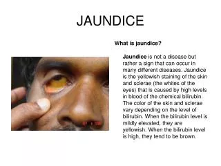
Introduction Condition where blockage of the flow of bile from the liver causes overspill of bile products into the blood and incomplete bile excretion from the body. More detailed information about the symptoms, causes, and treatments of Obstructive Jaundice is available below.
What are the causes of Jaundice? Some of the possible causes of Obstructive Jaundice include: Gallstones - most common cause Pancreatic cancer Hepatitis Drugs/medications Interstitial liver diseases
What are the symptoms of Obstructive Jaundice ? Some of the symptoms of Obstructive Jaundice include: Dark coloured urine Pale stools Yellow colouration of skin and eyes Itchy skin Fever
What treatments are available for Obstructive Jaundice ? Surgical removal of obstruction - generally keyhole (laparascopic) surgery or ERCP Cease drugs suspected to be causing liver inflammation - e.g. steroids, sulfonylureas Antibiotics Liver transplantation