Download
1 / 102
1.02k likes | 1.16k Views
Central Nervous System: “CNS”. Spinal Cord Brain. The Spinal Cord. Foramen magnum to L1 or L2 Runs through the vertebral canal of the vertebral column Functions Sensory and motor innervation of entire body inferior to the head through the spinal nerves

E N D
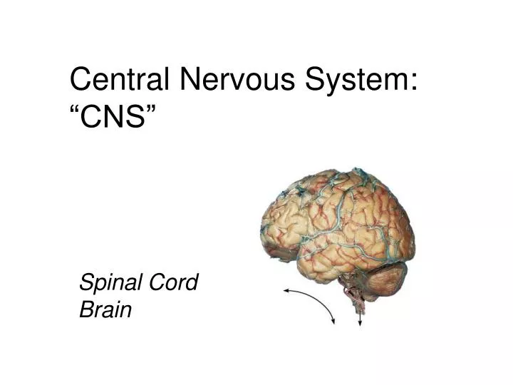
Central Nervous System:“CNS” Spinal Cord Brain
The Spinal Cord • Foramen magnum to L1 or L2 • Runs through the vertebral canal of the vertebral column • Functions • Sensory and motor innervation of entire body inferior to the head through the spinal nerves • Two-way conduction pathway between the body and the brain • Major center for reflexes
Spinal cord • Fetal 3rd month: ends at coccyx • Birth: ends at L3 • Adult position at approx L1-2 during childhood • End: conus medullaris • This tapers into filum terminale of connective tissue, tethered to coccyx • Spinal cord segments are superior to where their corresponding spinal nerves emerge through intervetebral foramina (see also fig 17.5, p 288) • Denticulate ligaments: lateral shelves of pia mater anchoring to dura (meninges: more later) http://www.apparelyzed.com/spinalcord.html
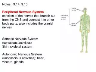
Spinal nerves • Part of the peripheral nervous system • 31 pairs attach through dorsal and ventral nerve roots • Lie in intervertebral foramina
Spinal nerves continued • Divided based on vertebral locations • 8 cervical • 12 thoracic • 5 lumbar • 5 sacral • 1 coccygeal • Cauda equina (“horse’s tail”): collection of nerve roots at inferior end of vertebral canal
Spinal nerves continued • Note: cervical spinal nerves exit from above the respective vertebra • Spinal nerve root 1 from above C1 • Spinal nerve root 2 from between C1 and C2, etc. • Clinically, for example when referring to disc impingement, both levels of vertebra mentioned, e.g. C6-7 disc impinging on root 7 • Symptoms usually indicate which level More about spinal nerves in the peripheral nervous system lecture
Bone Meninges CSF (cerebrospinal fluid) Protection: 3 meninges: dura mater (outer) arachnoid mater (middle) pia mater (inner) 3 potential spaces epidural: outside dura subdural: between dura & arachnoid subarachnoid: deep to arachnoid
Spinal cord coverings and spaces http://www.eorthopod.com/images/ContentImages/pm/pm_general_esi/pmp_general_esi_epidural_space.jpg • Dura mater • Arachnoid mater • Pia mater
LP (lumbar puncure) = spinal tap(needle introduced into subdural space to collect CSF) Lumbar spine needs to be flexed so can go between spinous processes Originally thought to be a narrowfluid-filled interval between the dural and arachnoid; now known to be an artificialspace created by the separation of the arachnoid from the dura as the result of trauma or some ongoing pathologicprocess; in the healthystate, the arachnoid is attached to the dura and a naturally occurring subdural space is not present. http://cancerweb.ncl.ac.uk/cgi-bin/omd?subdural+space • Epiduralspace is external to dura • Anesthestics are often injected into epidural space • Injection into correct space is vital; mistakes can be lethal
Spinal cord anatomy • Posterior median sulcus (“p”) • Anterior median fissure (“a”) • White matter (yellow here) • Gray matter (brown here) “p” “a”
Gray/White in spinal cord • Hollow central cavity (“central canal”) • Gray matter surrounds cavity • White matter surrounds gray matter (white: ascending and descending tracts of axons) • “H” shaped on cross section • Dorsal half of “H”: cell bodies of interneurons • Ventral half of “H”: cell bodies ofmotor neurons • No cortex (as in brain) Dorsal (posterior) white gray Central canal______ Ventral (anterior)
Spinal cord anatomy • Gray commissure with central canal • Columns of gray running the length of the spinal cord • Posterior (dorsal) horns (cell bodies of interneurons) • Anterior (ventral) horns (cell bodies of motor neurons) • Lateral horns in thoracic and superior lumbar cord * * * *
White matter of the spinal cord(myelinated and unmyelinated axons) • Ascending fibers: sensory information from sensory neurons of body up to brain • Descending fibers: motor instructions from brain to spinal cord • Stimulates contraction of body’s muscles • Stimumulates secretion from body’s glands • Commissural fibers: white-matter fibers crossing from one side of cord to the other • Most pathways cross (or decussate) at some point • Most synapse two or three times along the way, e.g. in brain stem, thalamus or other
The Brain: embryonic development • Develops from neural tube • Brain subdivides into • Forebrain • Midbrain • Hindbrain • These further divide, each with a fluid filled region: ventricle, aqueduct or canal • Spinal cord also has a canal • Two major bends, or flexures, occur (midbrain and cervical)
Brain development • Learn forebrain, midbrain and hindbrain in (b) • See next color coded pics in reference to (d) • Learn (e) • Encephalos means brain (otherwise you don’t need to learn “c”)
Space restrictions force cerebral hemispheres to grow posteriorly over rest of brain, enveloping it • Cerebral hemispheres grow into horseshoe shape (b and c) • Continued growth causes creases, folds and wrinkles
Anatomical classification • Cerebral hemispheres • Diencephalon • Thalamus • Hypothalamus • Brain stem • Midbrain • Pons • Medulla • Cerebellum • Spinal cord
Parts of Brain Cerebrum Diencephalon Brainstem Cerebellum
Usual pattern of gray/white in CNS • White exterior to gray • Gray surrounds hollow central cavity • Two regions with additional gray called “cortex” • Cerebrum: “cerebral cortex” • Cerebellum: “cerebellar cortex” _________________ ____________________________ _____________________________
Gray and White Matter • Like spinal cord but with another layer of gray outside the white • Called cortex • Cerebrum and cerebellum have • Inner gray: “brain nuclei” (not cell nuclei) • Clusters of cell bodies Remember, in PNS clusters of cell bodies were called “ganglia” More words: brains stem is caudal(toward tail) to the more rostral (noseward) cerebrum
Ventricles • Central cavities expanded • Filled with CSF (cerebrospinal fluid) • Lined by ependymal cells (these cells lining the choroid plexus make the CSF: see later slides) • Continuous with each other and central canal of spinal cord In the following slides, the ventricles are the parts colored blue
Lateral ventricles • Paired, horseshoe shape • In cerebral hemispheres • Anterior are close, separated only by thin Septum pellucidum
Third ventricle • In diencephalon • Connections • Interventricular foramen • Cerebral aqueduct
Fourth ventricle • In the brainstem • Dorsal to pons and top of medulla • Holes connect it with subarachnoid space
Subarachnoid space • Aqua blue in this pic • Under thick coverings of brain • Filled with CSF also • Red: choroid plexus (more later) ________
Surface anatomy • Gyri (plural of gyrus) • Elevated ridges • Entire surface • Grooves separate gyri • A sulcus is a shallow groove (plural, sulci) • Deeper grooves are fissures
Gyri (plural of gyrus) • Elevated ridges • Entire surface • Grooves separate gyri • A sulcus is a shallow groove (plural, sulci) • Deeper grooves are fissures
Parts of Brain Cerebrum Diencephalon Brainstem Cerebellum
simplified… • Back of brain: perception • Top of brain: movement • Front of brain: thinking
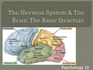
Cerebral hemispheres • Lobes: under bones of same name • Frontal • Parietal • Temporal • Occipital • Plus: Insula (buried deep in lateral sulcus)
Cerebral hemispheres: note lobes • Divided by longitudinal fissure into right & left sides • Central sulcus divides frontal from parietal lobes
Lateral sulcus separates temporal lobe from parietal lobe • Parieto-occipital sulcus divides occipital and parietal lobes (not seen from outside) • Transverse cerebral fissure separates cerebral hemispheres from cerebellum
coronal section • Note: longitudinal fissure, lateral sulcus, insula • Note: cerebral cortex (external sheet of gray), cerebral white, deep gray (basal ganglia)
Cerebral cortex • Executive functioning capability • Gray matter: of neuron cell bodies, dendrites, short unmyelinated axons • 100 billion neurons with average of 10,000 contacts each • No fiber tracts (would be white) • 2-4 mm thick (about 1/8 inch) • Brodmann areas (historical: 52 structurally different areas given #s) • Neuroimaging: functional organization (example later)
Prenatal life: genes are responsible for creating the architecture of the brain • Cortex is the last to develop and very immature at birth • Birth: excess of neurons but not inter-connected • 1st month of life: a million synapses/sec are made; this is genetic • 1st 3 years of life: synaptic overgrowth (connections) • After this the density remains constant though some grow, some die • Preadolescence: another increase in synaptic formation • Adolescence until 25: brain becomes a reconstruction site • Connections important for self-regulation (in prefrontal cortex) are being remodeled: important for a sense of wholeness • Causes personal turbulence • Susceptible to stress and toxins (like alcohol and drugs) during these years; affects the rest of one’s life • The mind changes the brain (throughout life) • Where brain activation occurs, synapses happen • When pay attention & focus mind, neural firing occurs and brain structure changes (synapses are formed) • Human connections impact neural connections (ongoing experiences and learning include the interpersonal ones) adapted from Dr. Daniel Siegel, UCLA
Cerebral cortex • All the neurons are interneurons • By definition confined to the CNS • They have to synapse somewhere before the info passes to the peripheral nerves • Three kinds of functional areas • Motor areas: movement • Sensory areas: perception • Association areas: integrate diverse information to enable purposeful action
Sensory areasPosterior to central sulcus • Primary somatosensory cortex: postcentral gyrus of parietal lobe (allows conscious awareness of sensation and the ability to localize it: where the sensation is from) • Somatosensory association area: behind it (understanding of what is being felt: the meaning of it)
From special sense organs • Sight: occipital lobe • Primary visual cortex (17) • Handles info from contralateral retina (right ½ of visual field is on left side) • Map of visual space • If damaged: functionally blind because no conscious awareness of sight • Visual association area (18 & 19) • Face recognition is usually on the right side • Hearing: temporal lobe • Primary auditory area (41) • Auditory association area (22)
Smell (olfactory sense): uncus • Deep in temporal lobe along medial surface
fMRI: functional magnetic resonance imaging • Cerebral cortex of person speaking & hearing • Activity (blood flow) in posterior frontal and superior temporal lobes respectively
Motor areas Anterior to central sulcus • Primary motor area • Precentral gyrus of frontal lobe (4) • Conscious or voluntary movement of skeletal muscles
Primary motor area continued • Precentral gyrus of frontal lobe • Precise, conscious or voluntary movement of skeletal muscles • Large neurons called pyramidal cells • Their axons: form massive pyramidal or corticospinal tracts • Decend through brain stem and spinal cord • Cross to contralateral (the other) side in brainstem • Therefore: right side of the brain controls the left side of the body, and the left side of the brain controls the right side of the body
Motor areas – continued • Broca’s area (44): specialized motor speech area • Base of precentral gyrus just above lateral sulcus in only one hemisphere, usually left • Word articulation: the movements necessary for speech • Damage: can understand but can’t speak; or if can still speak, words are right but difficult to understand
Motor areas – continued • Premotor cortex (6): complex movements asociated with highly processed sensory info; also planning of movements • Frontal eye fields (inferior 8): voluntary movements of eyes
Homunculus – “little man” • Body map: human body spatially represented • Where on cortex; upside down
Association Areas Remember… • Three kinds of functional areas (cerebrum) • Motor areas: movement • Sensory areas: perception • Association areas: everything else
Association Areas • Tie together different kinds of sensory input • Associate new input with memories • Is to be renamed “higher-order processing“ areas
Prefrontal cortex: cognition This area is remodeled during adolescence until the age of 25 and is very important for well-being; it coordinates the brain/body and inter-personal world as a whole Social skills Appreciating humor Conscience Mood Mental flexibility Empathy Intellect Abstract ideas Judgment Personality Impulse control Persistence Complex Reasoning Long-term planning Executive functioning e.g. multiple step problem solving requiring temporary storage of info (working memory)
Wernicke’s area Region involved in recognizing and understanding spoken words • Junction of parietal and temporal lobes • One hemisphere only, usually left • (Outlined by dashes) • Pathology: comprehension impaired for written and spoken language: output fluent and voluminous but incoherent (words understandable but don’t make sense; as opposed to the opposite with Broca’s area)