Download
1 / 93
930 likes | 946 Views
Immunology and Immunodeficiency. Sung-Yun Pai MD. Some points. Rest assured, this course (in my biased opinion) prepares you well for boards The content in this talk only ~5% of the questions Focuses on IMPORTANT cells of the blood—the LYMPHOCYTES!

E N D
Immunology and Immunodeficiency Sung-Yun Pai MD
Some points • Rest assured, this course (in my biased opinion) prepares you well for boards • The content in this talk only ~5% of the questions • Focuses on IMPORTANT cells of the blood—the LYMPHOCYTES! • Unfortunately the board questions seem to lag behind the content specs so some “old” content specs are included • * in the corner means there are some content specs on that slide • I have tinkered with this lecture in response to previous years’ feedback
Objectives • Identify the cellular basis of immune compromise in patients undergoing chemotherapy or HSCT, the classes of infections associated with various cellular deficiencies, and general principles of management of infections in immunocompromised hosts • Identify the normal pattern of development of the adaptive immune system in children and the presentation and diagnostic features of specific congenital deficiencies of adaptive immunity
Content to be reviewed 2 • Infections in the immunocompromised host • Normal T and B cell development and function and evaluation of recurrent infection • Congenital immunodeficiency states made easy • (2 slides on AIDS)
CD8 CD4 • Phagocytosis • Produce bactericidal reactive oxygen species neutrophils INNATE • Phagocytosis • Antigen presentation • Clearance of debris macrophages • Antigen presentation • Phagocytosis dendritic cells natural killer (NK) cells • Kill virally infected cells • Kill tumor or foreign cells CD16/ 56 ADAPT IVE • Cellular immunity • Help B cells class switch • Help CD8 T cells kill CD3/CD4 T cell thymus CD3/CD8 T cell • Cellular immunity • Cytolytic activity • Make antibody • Antibodies opsonize bacteria • Antigen presentation CD19 B cells 3
CD8 CD4 BACTERIA FUNGI neutrophils INNATE macrophages contribute to all dendritic cells VIRUSES (esp DNA viruses) natural killer (NK) cells CD16/ 56 ADAPT IVE CD3/CD4 T cell thymus VIRUSES PNEUMOCYSTIS FUNGI CD3/CD8 T cell BACTERIA (esp encapsulated) CD19 B cells 4
What host factors cause immunocompromise? • Loss of physical barriers • Medication • Acquired or congenital defects in cell number • Acquired or congenital defects in cell function 5
Chemotherapy and HSCT cause multiple hits to the immune system • Loss of physical barriers • mucosal impairment • central venous catheters etc. • Medication • corticosteroids (ALL, GVHD) • specific immunosuppressants (CsA, others) • antibody therapy (ATG, rituximab, alemtuzumab) • Acquired or congenital defects in cell number • neutropenia, CD4 lymphopenia • Acquired or congenital defects in cell function 6
* Timing of infections post-allogeneic HSCT Pre-engraftment (day 0-30) Post-engraftment (early day 30-100) Post-engraftment (late day >100) Gram + Gram - Anaerobes Same Esp catheter associated Encapsulated organisms* BACTERIA FUNGI Candida Aspergillus Aspergillus* Aspergillus* VIRUSES HSV Respiratory CMV* Respiratory EBV-LPD VZV CMV* Respiratory EBV-LPD OTHER Pneumocystis Toxoplasma Pneumocystis Toxoplasma DEFECT • profound neutropenia • catheter • lymphopenia esp CD4 • T cell suppressive meds • cGVHD • Poor Ig production • lymphopenia esp CD4 • T cell suppressive meds • aGVHD • catheter 7 * risk enhanced or associated with GVHD acute or chronic and its treatment
Management of infection in immunocompromised hosts Bacterial infection Fungal infection Pneumocystis Viral infection (especially DNA viruses) Note: these are not meant to be the absolute most up to date recommendations and are geared towards what I think are testable principles supported by evidence 8
* Bacterial infection In chemo/HSCT patients bacteria are derived from endogenous flora, in context of mucosal and skin barrier disruption Epidemiology has shifted from GNR to GPC GNR: E. coli, Pseudomonas, Enterobacter, Klebsiella, Serratia, other GPC: Staph aureus, coagulase negative Staph, alpha-hemolytic strep (S. mitis, S. sanguis), Enterococcus anaerobes: Bacteroides, Clostridium Association of Ara-C and alpha-hemolytic strep with ARDS and rapidly evolving sepsis syndrome Keep in mind resistant organisms and local pattern methicillin-resistant S. aureus, vancomycin-resistant enterococcus inducible plasmids encoding extended spectrum beta lactamases causing resistance after exposure to cephalosporins in Enterobacter and other GNR 9
* Bacterial infection Prophylaxis: Randomized controlled trials and meta-analyses demonstrate decrease in infection and possibly improved survival with prophylactic antibiotics in neutropenic cancer patients Benefits of use must be weighed against risk of developing resistance Principles of empiric therapy: Fever and neutropenia in setting of chemotherapy is an emergency Instituting immediate therapy without awaiting culture results is standard Physical examination to identify localizing source must be performed but rarely identifies cause Regimen should cover enteric GNR, S. aureus, other gram positives monotherapy is the standard recommendation (3rd gen cephalosporin, carbapenem, piperacillin/tazobactam) Additional agents (aminoglycoside, quinolone, more GPC coverage) upfront or delayed depending on clinical circumstances Stopping antibiotics after 7 days in persistently neutropenic patients leads to about 40% of patients developing fever or hypotension 10
Random interlude about the spleen * • Several immunological functions of the spleen • Site of filtration and innate immunity • Opsonization of encapsulated organisms • Consumption by macrophages • Site of primary and secondary adaptive immune responses • T cells get primed by APC • T cells interact with B cells, which make specific IgM • Activated follicular B cells mature, class switch, undergo affinity maturation • Site of specialized B cell population • Marginal zone B cells that respond to polysaccharide antigens and are generated by 2 years of age 11
Anatomic/functional asplenia * Prophylaxis: oral penicillin twice a day for <5 years old Lifelong for anyone who has had post-splenectomy sepsis Consider for >5 years old for first 1-2 years after splenectomy Principles of empiric therapy: Give antibiotics within 2 hours, at home if >2h from hospital For hospital: parenteral 3rd or greater generation cephalosporin For home: amoxicillin 45 mg/kg (max 2g) or levofloxacin 10 mg/kg (max 750 mg) Principles of vaccination: Special schedules for pneumococcus, HiB, meningococcus ACIP recommendations for immunocompromised (functional or anatomic asplenia, including sickle cell disease, HIV+, CSF leak, cochlear implant) 2012 Catch up everyone with PCV13 if PCV13 naïve Catch up everyone with PPSV23 if PPSV23 naïve If naïve to both, give PCV13 then 8 weeks later PPSV23 2nd dose of PPSV23 after 5 years If PPSV23 in past, but naïve to PCV13, give PCV13 >8 weeks from last PPSV23 12
* Fungal infection • Two major populations at risk, with propensity for different fungi • profound and long neutropenia • (Candida, Aspergillus, others) • defective cellular immunity • (Cryptococcus, Histoplasma, Coccidiodes) • Other rarer fungi include: • Fusarium, Mucor, Trichosporon, dematiaceous molds • Unlike bacterial infection, these are not endogenous but patients become colonized due to exposure and modulation of normal flora 13
* Fungal infection Prophylaxis: Large trials have shown decrease in fungal infections in adults during leukemia induction or undergoing HSCT Current IDSA recommendations are to use an azole (fluconazole, itraconazole, voriconazole, posaconazole) or echinocandin (micafungin, caspofungin) for prevention of Candidiasis during induction or HSCT (neutropenia >7 days) Patients with CGD benefit from prophylactic treatment Principles of empiric therapy: Empiric anti-fungal therapy is standard for persistent fever and neutropenia (4 to 7 days) Other possible signs of fungal disease include: nasal or sinus tenderness, painful swallowing, pulmonary infiltrates Therapy should be directed against Candida and Aspergillus, and no one agent is specifically recommended. Choice of empiric agent should take into account prophylactic regimen, known colonization, individual patient profile and local epidemiology 14
* Pneumocystis jirovecii (formerly called Pneumocystis carinii) risk factors for Pneumocystis pneumonia include: HIV infection with CD4 count <200 HSCT recipients cancer especially leukemia solid organ transplant recipients glucocorticoids, chemo or other immunosuppressants primary immunodeficiency especially SCID (renamed because P. jirovecii infects humans while P. carinii infects other mammals, like rodents) 15
* Pneumocystis jirovecii Prophylaxis: Trimethoprim-sulfamethoxazole 150 mg TMP/m2/day (5 mg/kg/day) divided BID TIW consecutive days most tested Alternatives to TMP/SMX: atovaquone, dapsone, aerosolized pentamidine, intravenous pentamidine, clindamycin plus primaquine, sulfadoxine plus pyrimethamine few if any studies in non-HIV patients comparing efficacy Treatment: Trimethoprim-sulfamethoxazole 15-20 mg/kg/day divided TID-QID, PO or IV for 14-21 days adjunctive corticosteroids shown to be effective in HIV, less certain in non-HIV Alternatives to TMP/SMX: mild disease: atovaquone, severe disease: pentamidine IV, clindamycin plus primaquine 16
* Viral infection • Patients with poor cell mediated immunity are at high risk for infection due to DNA viruses • post-BMT • solid organ transplant • HIV • primary immunodeficiency • CMV, HSV, VZV, EBV can all cause disease from primary exposure, or from reactivation of latent infection 17
* Viral infection Cytomegalovirus Herpes family DNA virus that causes asymptomatic or self-limited flu/mononucleosis illness in competent host Can be passed through organs, blood, breastmilk, saliva, urine Disease manifests as viremia, interstitial pneumonitis, colitis, retinitis, hepatitis, esophagitis/gastritis Asymptomatic shedding in urine and saliva can occur Organ disease and viremia/blood antigenemia are not always correlated Prophylaxis using only blood products from CMV negative donors OR leukofiltration in BMT setting, treatment with ganciclovir prevents interstitial pneumonia Treatment ganciclovir 5 mg/kg/dose IV twice a day for 2 weeks valganciclovir orally, dosing is not well established in children foscarnet 180 mg/kg/day divided twice or three times a day for 2 weeks cidofovir does have activity against CMV 18
* Viral infection Varicella zoster (Herpes zoster) Herpes family DNA virus that causes primary varicella and shingles Primary or reactivation disease can be severe in immunocompromised patients, causing hepatitis, pneumonitis, encephalitis Prophylaxis universal vaccination began in US in 1995 acyclovir, valacyclovir, famciclovir can all be used to prevent VZV reactivation in the absence of Varicella specific Ig (VZIG), pooled IVIg can be used to prevent disease after exposure in non-immune children up to 96h post Treatment high dose acyclovir, 1500 mg/m2/day IV divided every 8 hours, adjust for renal insufficiency and hydrate oral acyclovir or other anti-virals 19
Content to be reviewed 20 • Infections in the immunocompromised host • Normal T and B cell development and function and evaluation of recurrent infection • Congenital immunodeficiency states made easy • (2 slides on AIDS)
Lymphocytes contribute to both innate and adaptive immune systems * CD16/56 NK cell INNATE CD4 helper T cell thymus CD8 cytolytic (killer) T cell ADAPTIVE CD19 B cell 21
Major arms of the adaptive immune system * Cellular immunity T cells (most are a/b T cells) Express TCR with CD3 complex (TCRa, TCRb, CD3zz, CD3ed, CD3eg) -CD4 T cells see antigens complexed to MHC class II on APC helper function cooperate with B cells to induce class switching control opportunistic infection cancer surveillance regulatory functions -CD8 T cells see antigen complexed to MHC class I on APC cytolytic function kill virally infected cells control opportunistic infection cancer surveillance • Humoral immunity • B cells • Produce antibody • IgM and IgD first • IgG, IgA, IgE only after class switching • additional mutations in Ig locus occur—B cells carrying receptors with higher affinity are selected (affinity maturation) • Antibodies opsonize bacteria • particularly important for • respiratory pathogens • skin flora • encapsulated organisms 22
What holes are generated in the immune system when lymphocytes are absent or do not function? * Cellular immunity T cells -CD4 T cells loss of Ig production opportunistic infections fungal infections viral infections -CD8 T cells lack of killer function viral infection especially DNA viruses cancer surveillance Humoral immunity B cells Produce antibody Antibodies opsonize bacteria inability to opsonize bacteria especially respiratory, skin, encapsulated organisms 23
How does the adaptive immune system develop? PreT ProB PreB immB Blood Bone marrow Thymus TCR rearrangement CD3 TCR HSC HSC CD4 T T T BCR (Ig) rearrangement CD3 TCR proliferation preBCR CD8 BCR signaling T IgM Spleen CD19 IgM B IgD 24
How does the adaptive immune system adapt? PreB immB immB TCR TCR rearrangement CD4 Antigen T Helper function: B cell help T cell help T TCR HSC T CD8 Antigen proliferation T ProB Killer function: Lyse infected cells BCR (Ig) rearrangement IgM IgM CD19 Ig class switch Affinity maturation Antigen IgG B IgD IgA 25 Spleen & Lymph node IgM 25
How to measure immunologic function? * Enumeration total lymphocytes, subsets absolute lymphocyte count CD3 = T cells CD4 = helper T cells CD8 = cytolytic T cells CD19 = B cells CD16 or CD56 = natural killer (NK) cells 26
Normal values for lymphocyte subsets in children and adults * • ALC, CD3 and CD4 counts are higher in young children • B cell numbers are maximal 2 months to 2 years then contract • NK cell numbers are pretty stable 27
Infant versus adult lymphocyte subsets * Adult ALC 1800 (1000-2800) CD3 1200 (700-2100) CD4 700 (300-1400) CD8 400 (200-900) CD19 200 (100-500) CD56 300 (90-600) Infant (1 wk to 2 mo) ALC 6700 (3500-13100) CD3 4600 (2300-7000) CD4 3500 (1700-5300) CD8 1000 (400-1700) CD19 1000 (600-1900) CD56 500 (200-1400) ALC of less than 1000-2000 in an infant is highly abnormal. 28 values from Comans-Bitter J Peds 1997
How to measure immunologic function? * Non-specific function T cells Proliferation to mitogens phytohemagglutinin (PHA) pokeweed mitogen (PWM) concanavalin A (ConA) B cells Total IgG, IgA, IgM IgG subclasses IgG1, IgG2, IgG3, IgG4 Remember that antibody production also requires intact T cell function 29
* How to measure immunologic function? Specific function T cells Proliferation to antigen tetanus, Candida Skin testing Candida control • B cells • Antigen-specific antibody • protein Ag • (tetanus, hepBsAg) • carbohydrate antigen • (blood group, pneumovax) Remember that antibody production also requires intact T cell function 30
Immunoglobulins for dummies * Class Structure Affinity Placental transfer? % IgM Pentameric Low affinity No 5-10% IgG Monomeric High affinity Yes 75-85% IgA Mono-, dimeric Low affinity No 5-15% * IgD does not require class switching, no clear function * Levels of IgE not necessarily part of an immunodeficiency work-up 31
Normal immunoglobulin for age (IgG) * physiologic nadir 2-6 months 32
Take home points about normal lymphoid development * 35 • infants and children have higher ALC and T cell numbers than adults • IgG crosses the placenta • physiologic nadir of IgG occurs around 2-6 months
Content to be reviewed 36 • Infections in the immunocompromised host • Normal T and B cell development and function and evaluation of recurrent infection • Congenital immunodeficiency states made easy • (2 slides on AIDS)
Congenital Immunodeficiency in one slide * AbsentPresent/Broken T cells B cells Neutrophils SCID Ex: WAS XLA Ex: CVID SCN Ex: CGD 37 Note: patients lacking NK cells have been described but are very rare
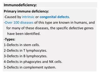
Congenital Immunodeficiency * 38 • Failure of T cell development SCID (severe combined immunodeficiency) • Failure of B cell development XLA (X-linked agammaglobulinemia) • Functional T, B, or combined defects T and B cells present but not fully functional Wiskott-Aldrich syndrome (WAS) X-linked Hyper-IgM syndrome (CD40LG) X-linked lymphoproliferative disease (XLP) Common variable immunodeficiency (CVID) Autoimmune lymphoproliferative syndrome (ALPS)
Congenital Immunodeficiency * 39 • Failure of T cell development SCID (severe combined immunodeficiency) • Failure of B cell development XLA (X-linked agammaglobulinemia) • Functional T, B, or combined defects T and B cells present but not fully functional Wiskott-Aldrich syndrome (WAS) X-linked Hyper-IgM syndrome (CD40LG) X-linked lymphoproliferative disease (XLP) Common variable immunodeficiency (CVID) Autoimmune lymphoproliferative syndrome (ALPS)
Definition of SCID * 40 “genetically heterogeneous group of syndromes having in common a profound disturbance in the development and function of both T and B cells” • Absence of functional autologous T cells is uniform • B cells can be present or absent • B cells even if present usually don’t function, i.e. no specific antibody production • generally fatal before age 2 due to overwhelming infection
When to suspect SCID * • Symptoms • Failure to thrive • Chronic diarrhea • Recurrent infections • Signs/Lab findings • thrush • absence of lymphoid tissue • opportunistic infection ex: PCP • low or absent T cells • lack of immunoglobulins • lack of thymic shadow • Associated findings • FH ex: X-linked • neurologic (ADA) • cardiac (DiGeorge) 41
How to diagnose SCID * Check ALC, send lymphocyte subsets T cells very low or absent Send lymphocyte proliferation studies Absent or very low proliferation of T cells to mitogens Total IgG, IgA, IgM and specific Ab if vaccinated 42
SCID with normal ALC? SCID with T cells present? * 4 month old boy with chronic cough and FTT. Diagnosed with PCP. CBC: WBC 12 60% polys 35% lymphs ANC = 7200 ALC = 4200 (3700-9600) CD3 = 400 (2300-6500) CD4 = 8 (1500-5000) CD8 = 300 (500-1600) CD19 = 3595 (600-3000) CD56= 15 (100-1300) FISH XX/XY = 2% XX proliferation to mitogens: flat IgG 87 (196-558) 43
How to diagnose SCID * Check ALC, send lymphocyte subsets Because B cells can be present, normal ALC does not rule out SCID Because maternal engraftment can occur, presence of T cells does not rule out SCID Send lymphocyte proliferation studies Maternal T cells do not proliferate. Normal proliferation rules out classic SCID. Total IgG, IgA, IgM and specific Ab if vaccinated Molecular defect can be narrowed down based on lymphocyte phenotype: T-B+ (can be NK - or +) T-B- (can be NK - or +) 44
T- B+ SCID * Molecular defects: cytokine common gamma chain (IL2RG, c, CD132) JAK3 (JAK3, Janus kinase 3) IL-7 receptor alpha chain (IL7R, IL-7R, CD127) B cells are present but receive no help, generally do not produce antibody c has X-linked inheritance, accounts for about 1/3 of all SCID JAK3 and IL-7R are autosomal recessive 45
Lack of growth factor signaling leads to absence of T cells IL-4 Jak3 * c IL-7R IL-2 IL-7 Jak3 c c Jak3 Jak3 c Jak3 IL-15 c IL-21 Jak3 c IL-9 46
Normal adaptive immune system development ProB PreB PreT immB Blood Bone marrow Thymus TCR rearrangement CD3 TCR HSC HSC CD4 T T T BCR (Ig) rearrangement CD3 TCR proliferation preBCR CD8 BCR signaling T IgM Spleen CD19 IgM B IgD 47
Lack of cytokine signaling causes loss of T cells PreB ProB immB Blood Bone marrow Thymus TCR rearrangement HSC HSC BCR (Ig) rearrangement X proliferation preBCR BCR signaling IgM Spleen CD19 IgM B IgD 48