Download
1 / 53
560 likes | 994 Views
Immunodeficiency Immunology of Pregnancy Immunity in the Elderly. Lecture objectives are to understand: How do microbial pathogens evade the immune system? Genetic disorders that cause immunodeficiency How does AIDS virus evade the immune system and cause immuodeficiency?

E N D
Immunodeficiency Immunology of Pregnancy Immunity in the Elderly
Lecture objectives are to understand: • How do microbial pathogens evade the immune system? • Genetic disorders that cause immunodeficiency • How does AIDS virus evade the immune system and cause immuodeficiency? • Why does the immune system become weaker as we get older? • Why are fetuses not rejected in pregnant mothers despite their MHC differences?
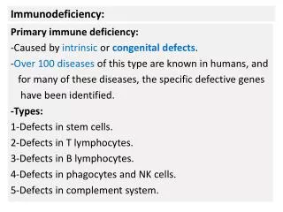
Why do we fail to mount effective immune response to pathogens? • 1. Congenital immunodeficiency • (defective immune cells or molecules of the immune system) • 2. Acquired immunodeficiency by Human Immunodeficiency Virus • 3. Pathogens can evade the immune system • (e.g. multiple serotypes, mutations, gene conversion, hiding in dormant state, blocking immune response, and superantigens)
CORE 24. Immunodeficiency Diseases • General signs and symptoms of immunodeficiency diseases Usually patients suffer from chronic infections of opportunistic pathogens High incidence of cancer
You are immune-deficient if defective in any of the following components of the immune system • Signaling molecules in TCR activation • Key transcriptional factors of immune cells • Molecules and organs for lymphocyte development • Antigen presentation • Phagocyte function • Cytokines and co-stimulation molecules • Immune cell migration and adhesion • Complement • DNA recombination and metabolism
CORE b. Laboratory tests to assess immune function (1) T cell: Enumeration (flow cytometry), functional assays (mitogen response, MLR, DTH skin tests) (2) B cell: Enumeration, circulating antibody levels (3) Macrophage: Enumeration, functional assays (nitroblue tetrazolium) (4) Complement: Direct measurement of complement components, complement hemolysis assay
CORE • Primary B cell immunodeficiencies (symptoms, description of defects, current therapy) • (1) X-linked Agammaglobulinemia (Bruton's syndrome); btk deficiency • (2) Common Variable Immunodeficiency (acquired hypogammaglobulinemia) • (3) Selective IgA deficiency (most common immunodeficiency disorder) • (4) Other (minor): • (a) Transient hypogammaglobulinemia of infancy • (b) Selective deficiency of IgG subclasses • (c) Immunodeficiency with hyper IgM
CORE • Primary T cell immunodeficiencies (symptoms, description of defect, current therapy) (1) Congenital thymic aplasia (DiGeorge's Syndrome or Third and Fourth Pharyngeal Arch Syndrome)
CORE • e. Combined B and T cell immunodeficiencies (symptoms, description of defect, current therapy) • Severe Combined Immunodeficiency (SCID; a group of genetically determined diseases) • X-Linked combined immunodeficiency (accounts for 50-60% of all SCID; defect in cytokine receptors) • Adenosine deaminase deficiency (an autosomal recessive SCID; accounts for ~20% of all SCID) • (c) Other mechanisms of SCIDs: Purine nucleoside phosphorylase deficiency, TCR immunodeficiency, MHC class I or II deficiency (Bare Lymphocyte Syndrome), Defective IL-2 production
Figure 9-7 part 1 of 3 Inherited immunodeficiencies Bubble boy =SCID =SCID BLS =SCID
Caused by deficiency in common gamma chain (γc) : The Cytokine receptor common gamma chain (γc) (or CD132) is a cytokine receptor sub-unit that is common to the receptor complexes for at least six different interleukin receptors: IL-2, IL-4, IL-7, IL-9, IL-15 and interleukin-21 receptor.
Figure 9-7 part 2 of 3 Inherited immunodeficiencies Most common
Figure 9-8 X-linked agammaglobulinemia Mutation in the btk gene Bruton's agammaglobulinemia tyrosine kinase (btk). Failure in B cell development Males are more affected
Figure 9-7 part 3 of 3 Other inherited immunodeficiencies
CORE f. Primary phagocyte deficiencies (symptoms, description of defect, current therapy) • (1) Neutropenia • (2) Chronic Granulomatous Disease • (3) Leukocyte Adhesion Deficiency
Figure 9-10 Defects in phagocytic cells: persistent bacterial infections
CORE • Primary complement deficiencies (symptoms, description of defect, current therapy) • (1) Deficiency of Complement Components • (a) Classic pathway: C1, C4, C2, C3 • (b) Alternative pathway: Factor D, Properdin • (c) MAC: C5, C6, C7, C8, C9 • (e) Regulator proteins: Factors H, I, C1 inhibitor • Hereditary Angioedema (C1INH deficiency)
Deficiencies in the pathways of complement activation Clearance of immune (Ab-Ag) complex Opsonin Membrane attack Enhances alternative path Supplies C3 Prevent host cell destruction
Bone marrow transplantation is used to correct genetic defects of the immune system High degree of HLA matching between patients and donors are critical: • To prevent alloreactions causing GVHD (graft-vs-host disease) and graft rejection • For effective presentation of antigen to donor-derived T cells
CORE • Secondary immunodeficiencies • Drug or radiation-induced (steroids, other cytotoxic drugs) • AIDS (HIV target cells and immune dysfunction [see Virology for other aspects of the viral infection]) • Nutritional deficiency (reduced protein, calorie, biotin, B12, Iron, Vit. A, Zinc; thymic atrophy pathologic result) • Autoimmune Disease (?, frequent inflammatory diseases are found in immunodeficient patients) (5) Other (postviral, chronic infection, neoplastic diseases)
Figure 9-13 Human Immunodeficiency Virus
Progression of AIDS Infection viremia (increase of the virus load in blood) immune response to HIV: generation of Tc cells and antibody to HIV (seroconversion) temporary reduction of virus-infected CD4 T cells (due to HIV-induced apoptosis and T cell attack) partial recovery of CD4 T cell number gradual decrease of CD4 T cell number over 2-15 years (clinical latency is a period of active infection and CD4 T cell renewal) AIDS (CD4 T cell count < 200)
Acquired Immune deficiency syndrome • HIV infection through the host CD4 molecule and chemokine receptors (CCR5 or CXCR4) • T cell activation is required for viral replication • Initial viremia and CD4 T cell number decrease (asymptomatic or mild flu-like) • Anti-HIV response and CD4 T cell number recovery • Long clinical latency (2- 15 yrs): a period of active viral infection and CD4 T cell renewal; gradual CD4 T cell number decrease • Immunodeficiency starts when the CD4 count is below 500 Opportunistic infection • Death due to secondary infection
HIV receptor and coreceptors • HIV receptor: CD4 • Co-receptors: chemokine receptors CCR5 and CXCR4 • Macrophage-tropic virus: infects macrophages, DCs and some T cells through CCR5 (early stage virus) • Lymphocyte or T-tropic HIV infects T cells through CXCR4 (late stage virus). • Switch from M-tropic to T-tropic occurs in 50% of cases. • HIV replication requires T cell activation (NF-Kb for viral RNA transcription)
Figure 9-15 The life cycle of Human Immunodeficiency Virus
Gradual loss of CD4 T cells after infection • There are long-term non-progressors; some remain seronegative • Some people have mutant CCR5 thus can be resistant to HIV
Figure 9-20 Combination drugs are effective in reducing HIV in patients However this is not complete elimination
Figure 9-22 Opportunistic infections and malignancies kill HIV patients
HIV kills CD4 T cells CD40L IL-4, IL-5, IL-10 and IFN-g Ag-presentation & activate IFN-g NK Killing of HIV+ cells Killing of HIV+ cells Anti-HIV antibody Immune cells involved in anti-HIV response Adaptive Immunity Innate Immunity NK DC CD4 CD8 B
HIV: why is it formidable? • High mutability and slow progression: • Changes in viral antigens (immune targets); • and enzymes (drug targets) • Generation of viral variants • Difficult to make vaccines • High rates of vertical and lateral transmissions • Targets CD4 T cells • One HIV drug is effective transiently. • Combination therapy using multiple drugs are more effective.
HIV evades host immune response by rapid mutation • HIV and other retroviruses have error-prone reverse transcriptases, generating mutant variant viruses or quasi-species • Selection of mutant viruses that have lost the epitope recognized by the host immune system • Sometimes, homologous peptides of variant viruses interfere the presentation of the original peptides • Such diversity greatly complicates vaccine development and limits the effectiveness of anti-viral drugs
Figure 9-1 Genetic variations within some species of pathogens prevent effective long-term immunity: 90 serotypes of Streptococcus pneumonia
Mutation and recombination allow influenza virus to escape from immunity RNA virus has genome of 8 RNA molecules Ag: Neuraminidase and Hemagglutinin Mutation leads to antigen drift: mild and limited epidemics Recombination or swapping RNAs leads to antigen shift: severe pandemics, every 10-50 years
Unicellular protozoa that are parasites of insects, plants, birds, bats, fish, amphibians and mammals. Trypanosomes undergo gene conversion to change their surface antigens African trypanosomes causes sleeping sickness, transmitted through insect bites, and have 1000 copies of inactive Variable Surface glycoprotein genes. Chronic cycle of antibody production and antigen clearance leads to a heavy deposition of immune complexes, inflammation, neurological damages and finally coma. Another example is the pilin change by Neisseria gonorrheae Salmonella tryphimurium alternates expression of two flagellins
Herpes virus persists in human hosts by hiding from the immune response in a dormant state Herpes simplex virus causes cold sores, first infect epithelial cells and then spreads to sensory neurons. Viruses persist in a latent state in the sensory neurons. Various stresses reactivate the virus. Reactivated viruses infect epithelial cells. Neuron is a favored place to hide because it expresses low levels of MHC class I molecules. Other examples are: the herpes virus varicella-zoster causing shingles hides in ganglia and Epstein-Barr virus causing mononucleosis hides in B cells.
Figure 9-5 part 1 of 3 Mechanisms of viruses to subvert the immune system
Figure 9-5 part 2 of 3 Mechanisms of viruses to subvert the immune system
Figure 9-5 part 3 of 3 Mechanisms of viruses to subvert the immune system
Bacterial pathogens subvert the immune system • M. tuberculosis: prevents fusion of phagosomes with lysosomes • Listeria monocytgenes: escapes phagosomes • Treponema pallidum: coats itself with human proteins • Staphylococci: produces superantigens (SEB and TSST-1) • Mycobacterium leprae: induces Th2 response to evade Th1 CMI response
Figure 9-6 Superantigens induce polyclonal T cell activation, inhibiting antigen-specific immunity
CORE 26. Immunity in the Elderly • a. Effects of aging on the immune system; thymic atrophy
CORE 25. Immunology of Pregnancy • Mucosal immunity of the female genital tract (IgA secretion) • Trophoblasts and immune responses Trophoblasts don’t express MHC I and II (reduced immunogenicity of fetal cells) Active immune suppression by secretion of immunosuppressive factors such as α-fetoprotein,IL-10 and TGF-β Inhibition by T-regulatory/suppressor cells
In pregnancy, the endometrium represents the hosting surface for invading semi-allogeneic cells (trophoblasts). The immunological mechanism must provide a balanced environment whereby the conceptus is nurtured by the mother and yet prevented from excessive invasion. Decidual natural killer cells play a role in the implantation and early invasion of the trophoblast. Regulatory T cells in suppression of immune responses against fetus (?)
Immunological factors leading to pregnancy failure Stages of normal pregnancy Gamete Development Fertilization Embryo Cleavage Trophoblast formation Implantation Fetal Development Parturition Anti-sperm Ab Defective adhesion molecules Anti-phospholipid Ab Aberrant HLA expression NK cell attack (inhibited by HLA-G) Too much Th1 cytokine? T cell attack Lack of suppressor T cells
1. RhD- mother can reject RhD+ fetus at second pregnancy. Hemolytic disease of the fetus and newborn Anti-RhD (IgG) antibodies can cross placenta. Administration of anti-RhD antibodies (Rhesus prophylaxis) either during pregnancy or immediately after delivery can prevent it. 2. Why does not a blood type O mother reject A type fetus? Anti-RhA or B (IgM) antibodies can not cross placenta. 3. An RhD−/type O mother carrying an RhD+/type A, B, or AB fetus is resistant to sensitization to the RhD antigen . No hemolytic disease in this case. Anti-A/B clears fetal cells in the mom.