Download
1 / 1
20 likes | 138 Views
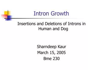
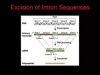
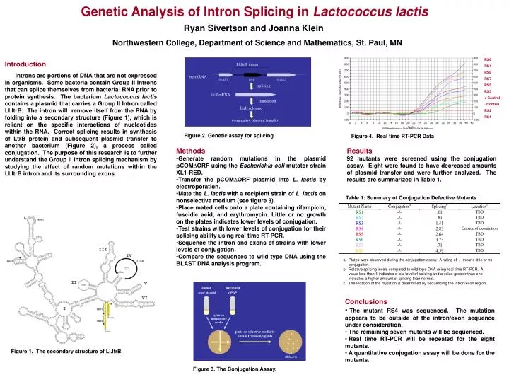
Ll.ltrB intron. pre-mRNA. ltrBE1. ltrA. ltrBE2. splicing. ltrB mRNA. translation. LtrB relaxase. conjugative plasmid transfer. Donor. Recipient. rif R fa R. erm R plasmid. grow on nonselective media. plate on selective media to obtain transconjugants. rif,fa,erm.

E N D
Ll.ltrBintron pre-mRNA ltrBE1 ltrA ltrBE2 splicing ltrB mRNA translation LtrB relaxase conjugative plasmid transfer Donor Recipient rifRfaR ermR plasmid grow on nonselectivemedia plate on selective media to obtain transconjugants rif,fa,erm Genetic Analysis of Intron Splicing in Lactococcus lactis Ryan Sivertson and Joanna Klein Northwestern College, Department of Science and Mathematics, St. Paul, MN • RS6 • RS4 • RS8 • RS7 • RS5 • RS3 • + Control • Control • RS2 • RS1 Introduction Introns are portions of DNA that are not expressed in organisms. Some bacteria contain Group II Introns that can splice themselves from bacterial RNA prior to protein synthesis. The bacterium Lactococcus lactis contains a plasmid that carries a Group II Intron called Ll.ltrB. The intron will remove itself from the RNA by folding into a secondary structure (Figure 1), which is reliant on the specific interactions of nucleotides within the RNA. Correct splicing results in synthesis of LtrB protein and subsequent plasmid transfer to another bacterium (Figure 2), a process called conjugation. The purpose of this research is to further understand the Group II Intron splicing mechanism by studying the effect of random mutations within the Ll.ltrB intron and its surrounding exons. Figure 2. Genetic assay for splicing. Figure 4. Real time RT-PCR Data • Methods • Generate random mutations in the plasmid pCOM∆ORF using the Escherichia coli mutator strain XL1-RED. • Transfer the pCOM∆ORF plasmid into L. lactis by electroporation. • Mate the L. lactis with a recipient strain of L. lactis on nonselective medium (see figure 3). • Place mated cells onto a plate containing rifampicin, fuscidic acid, and erythromycin. Little or no growth on the plates indicates lower levels of conjugation. • Test strains with lower levels of conjugation for their splicing ability using real time RT-PCR. • Sequence the intron and exons of strains with lower levels of conjugation. • Compare the sequences to wild type DNA using the BLAST DNA analysis program. Results 92 mutants were screened using the conjugation assay. Eight were found to have decreased amounts of plasmid transfer and were further analyzed. The results are summarized in Table 1. Table 1: Summary of Conjugation Defective Mutants • Plates were observed during the conjugation assay. A rating of -/- means little or no conjugation. • Relative splicing levels compared to wild type DNA using real time RT-PCR. A value less than 1 indicates a low level of splicing and a value greater than one indicates a higher amount of splicing than normal. • The location of the mutation is determined by sequencing the intron/exon region. • Conclusions • The mutant RS4 was sequenced. The mutation appears to be outside of the intron/exon sequence under consideration. • The remaining seven mutants will be sequenced. • Real time RT-PCR will be repeated for the eight mutants. • A quantitative conjugation assay will be done for the mutants. Figure 1. The secondary structure of Ll.ltrB. Figure 3. The Conjugation Assay.