Download
1 / 7
80 likes | 244 Views
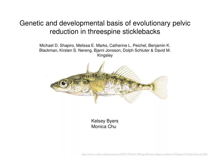
Genetic and developmental basis of evolutionary pelvic reduction in threespine sticklebacks. Michael D. Shapiro, Melissa E. Marks, Catherine L. Peichel, Benjamin K. Blackman, Kirsten S. Nereng, Bjarni Jonsson, Dolph Schluter & David M. Kingsley. Kelsey Byers Monica Chu.

E N D
Genetic and developmental basis of evolutionary pelvic reduction in threespine sticklebacks Michael D. Shapiro, Melissa E. Marks, Catherine L. Peichel, Benjamin K. Blackman, Kirsten S. Nereng, Bjarni Jonsson, Dolph Schluter& David M. Kingsley Kelsey Byers Monica Chu http://www.colby.edu/chemistry/CH217/Fish%20Page/Pictures/Species/three%20spine%20stickleback.JPG
Vertebrate Limb Structures • Highly varied • In phylogenetically diverse sets of vertebrates, hindlimb structures are reduced or completely absent • Little is known about the mechanisms underlying the genetic changes that lead to hindlimb reduction in vertebrate evolution • Marine threespine sticklebacks • - prominent pelvic skeleton • - protection against predators • Freshwater threespine sticklebacks- reduced or complete loss of pelvic structures
F1 Marine stickleback Benthic Paxton Lake Stickleback F2 375 F2 progeny (289 unaffected : 86 affected) mednews.stanford.edu/story_images/stickleback.jpg
Table 1 • One major QTL on linkage group 7 • Four minor QTL on linkage groups 1, 2, 4, and 10 • Region near linkage group 7 accounts for a large amount of variance in pelvic • structure • Presence of freshwater alleles decreased the average size of pelvic structures
Figure 1 Increasing the total number of benthic alleles at the minor QTL of linkage groups 1, 2 and 4 resulted in greater reduction of pelvic structures than the effects of a minor QTL alone
Figure 1 • Tbx4 gene mapped to linkage group 1 • Pitx2 gene mapped to linkage group 4 • Pitx1 gene mapped to the area that seems to be the major controlling region for pelvic reduction
Left-right assymetry in stickleback pelvic reduction • Pitx1 gene in the mouse is responsible for hindlimb development • Pitx1 knockout mice exhibit strong directional assymetry • Directional assymetry observed in F2 progeny • -stronger development on the left side in 78% of fish with pelvic assymetry