Download
1 / 12
170 likes | 542 Views
The Hip Joint. Type: Synovial (Ball & Socket) Articular Surfaces: head of femur & acetabulum. Articular surfaces: 1- Acetabulum has: - C- shaped articular surface = lunate surface, Acetabular fossa,

E N D
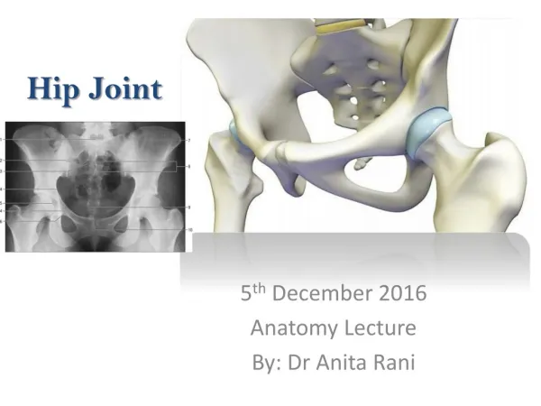
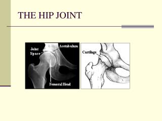
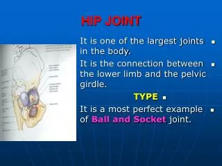
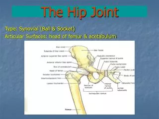
The Hip Joint Type: Synovial (Ball & Socket) Articular Surfaces: head of femur & acetabulum
Articular surfaces: 1- Acetabulumhas: - C- shaped articular surface = lunate surface, Acetabular fossa, • Acetabular notch -----► converted into acetabular foramen by transverse ligament of the acetabulum • - Labrum acetabulare deepens the acetabular cavity. • 2- Head of femur
Ligaments: 1- Ilio-femoral lig.: Y- shaped, strong, attached between AIIS and both ends of trochantric line. Prevents hyper-extension. 2- Pubo-femoral lig.:Triangular in shape, supports the inferomedial part of the joint, attached between ilio-pubic eminence and the capsule. Prevents hyper-abduction. 3- Iscio-femoral lig.: from ischium to the back of the capsule. It becomes tense in medial rotation. 4- round ligament of the head of femur: Inside the joint, between head of femur to acetabular notch and transverse lig., carries blood supply to head of femur.
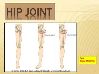
Movements: 1- Flexion: Psoas major, Iliacus + Rectus femoris, sartorius, pectineus. 2- Extension: Gluteus maximus + hamstring 3- Abduction: Gluteus medius & minimus + tensor fascia latae 4- Adduction: Adductor longus, brevis, magnus + gracilis, pectineus 5- Medial rotation: Gluteus medius & minimus + tensor fascia latae 6- Lateral rotation: The 6 lateral rotators: Obturator internus, 2 gemelli, piriformis, obturator externus, quadratus femoris + gluteus maximus, adductors. 7- Circumduction: Nerve supply of hip joint: 1- Femoral nerve. 2- Obturator nerve. 3- sciatic nerve. 4- Nerve to quadratus femoris.
Relations of the hip joint: • Anteriorly: Pectineus, Iliopsoas, RF (straight head), femoral vessels. • Laterally: Tensor fascia latae, gluteus minimus & medius. • Posteriorly: Piriformis, obturator internus, 2 gemelli, quadratus femoris, sciatic nerve. • Above: RF (reflected head), gluteus minimus. • Below: Obturator externus.
Stability of the hip joint: The hip joint is very stable joint due to: 1- The head of femur fits accurately to the acetablum. 2- The three strong ligaments outside the capsule. 3- The surrounding strong muscles. Hip dislocation is usually posterior as in car accidents. It occurs with no fracture of the acetabulum (if the hip is flexed and adducted) or with fracture acetabulum (if the hip is flexed and abducted). The sciatic nerve may be injured in posterior hip dislocation.
Blood supply to acetabular fossa and ligament of the head of femur: