Download
1 / 40
400 likes | 479 Views
Explore the structure of neurons, spinal cord, brain, and sensory organs in detail with labeled diagrams and descriptions. Understand reflex arcs, nervous system functions, and sensory experiments. Learn about cranial nerves, peripheral nerves, and eye and ear anatomy.

E N D
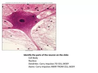
Identify the parts of the neuron on the slide: Cell Body Nucleus Dendrites- Carry impulses TO CELL BODY Axons- Carry impulses AWAY FROM CELL BODY
Dura Mater- outter mostArachnoid Mater- middlePiaMater- innermost
3. Know the parts of the Spinal Cord Model Posterior Median Sulcus Anterior Median Fissure Central Canal – filled with CSF Gray Matter- Posterior horn Lateral horn Anterior horn Grey commisure- connects two halves of gray matter White Matter- Poteriorfuniculus Lateral funiculus Anterior funiculus Spinal Nerve Doral root- carries sensory impulse INTO spinal cord (afferent) Dorsal root ganglion- contains SENSORY NEURON CELL BODIES Ventral root- carries motor impulse AWAY from spinal cord (efferent)
4. Know the Periphereal Nerves Peripheral Nerves ARM: Radial - outside Ulnar - inside Median –middle
LEG: Sciatic – can only see on the left foot Obturator- pass through the foramen Femoral- runs down the femur
Know the parts of the brain model Cerebrum Cerebral hemispheres seperated by LONGIDUTINAL FISSURE Corpus Callosum- connects the 2 cerebral hemispheres Gyri- Precentral – motor area Postcentral – sensory area Sulci- Central Sulcus Lateral Sulcus Central Lobes- Frontal Parietal Temporal Occipital Broca’s Area- Motor speech area
Diencephalon Thalamus – gateway for sensory impulse (except smell) Hypothalamus- PITUITARY GLAND and PITUITARY STALK Pineal Gland – Melatonin Cerebellum Transverse fissure – separate cerebrum from cerebellum Arbor Vitae- pattern of white matter in cerebellum Brain Stem Midbrain Pons Medulla Oblongata
Know the Cranial Nerves • Olfactory- Smell • Optic – Vision • Oculomotor- 4 eye muscles • Trochlear- 1 eye muscle • Trigeminal- sensory nerve of face • Abducens- 1 eye muscle • Facial- muscles for facial expressions • Auditory- equilibrium and hearing • Glossopharyngeal- innervates muscle of throat • Vagus- All major organs • Accesory- muscles of neck and shoulder • Hypoglossal- muscle of tongue
Know the reflex arc • Receptor • Sensory Neuron • Inner Neuron • Motor Neuron • Effector
Experiments • Two point discrimination test – determines the density of touch receptors in different areas of the body.
Pressure sense acuteness – determine the distribution of touch receptors
Adaptation of Touch Receptors – number and strength of sensory impulses changes overt time
Referred Pain – where pain is felt in one part of the body when the stimulus is elsewhere
Experiments • Localization of Taste – mapping of the taste buds • Hearing tests Rinne’s – detects conduction deafness Weber’s – detects nerve deafness
Visual Acuity- 20/20 1st number is your eye, 2nd number is healthy eye
Blind Spot Determination- The blind spot is created by the area where the optic nerve connects to the retina.
Photopupillary Reflex- Whenever the light shines in the eye the iris constricts
Negative Color After Image – resynthesize the pigments opposites
Know the anatomy of the ear Auricle External Auditory Meatus Tympanic Cavity holds Malleus Incus Stapes Tympanic Membrane Auditory Tube Oval Window Vestibule Semicircular canal Cochlea Round Window Auditory Nerve (Vestibulocochlear)
Know the anatomy of the Eye Eyelid Lacrimal Gland Lacrimal Sac MUSCLES OF THE EYE Obicularis Oculi- surrounds eye Superior Rectus – rolls eye up Inferior Rectus- rolls eye down Lateral Rectus- rolls eye outward Medial Rectus- rolls eye inward Superior Oblique- rolls eye down Inferior Oblique- rolls eye up
Outer tunic (layer) Sclera- white Cornea- transparent Middle Tunic (layer) Choroid coat- dark Ciliary body with ciliary muscles Suspensory ligaments Lens- transparent Iris- color varies Pupil- hole in iris, allows light waves to reach retina, size of pupil determined by iris Inner Tunic (layer) Retina- contains photoreceptors called RODS and CONES RODS detect light, CONES detect color
Anterior cavity- located in front of the lens and contains AQUEOUS HUMOR Anterior chamber- between cornea and iris Posterior chamber- between iris and lens Posterior cavity- located between lens and retina contains VITREOUS HUMOR