Download
1 / 69
810 likes | 1.15k Views
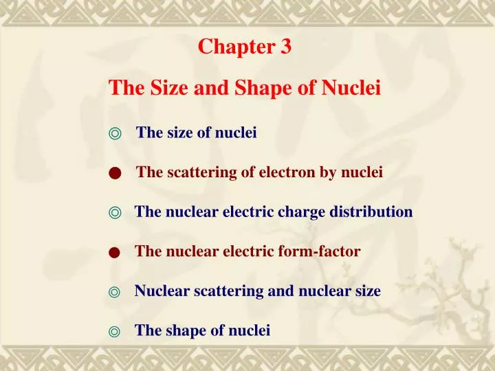
Chapter 3 The Size and Shape of Nuclei. ◎ The size of nuclei ● The scattering of electron by nuclei ◎ The nuclear electric charge distribution ● The nuclear electric form-factor ◎ Nuclear scattering and nuclear size ◎ The shape of nuclei.

E N D
Chapter 3 The Size and Shape of Nuclei ◎The size of nuclei ●The scattering of electron by nuclei ◎The nuclear electric charge distribution ●The nuclear electric form-factor ◎Nuclear scattering and nuclear size ◎The shape of nuclei
“7And I will establish My covenant between Me and you and your seed after you throughout their generations for an everlasting covenant, to be God to you and to your seed after you.8And I will give you and to your seed after you the land of your sojournings, all the land of Canaan, for an everlasting possession; and I will be their God.” (Genesis 17:7 ~ 8)
§ 3.1 The size of nuclei • What we are interested here is • the size of nuclei • how the nuclear matter is distributed inside a sphere, if the nucleus • is spherical. In fact Nuclei are not always spherical! A prolate deformed nucleus
What we mean by the size of a nucleus ? Rough visualization of an electron "wave function". (Art by Blake Stacey.) The electron probability density for the first few hydrogen atom electron orbitals shown as cross-sections. These orbitals form an orthonormal basis for the wave function of the electron. In atomic physics, the boundary of an atom is not sharp since the wave function of the outer electrons decreases monotonically. We have a similar situation in the nuclear physics!
These deviations occur at short distances of approach. • the finite charge density distributions overlap. • 2 strong nuclear forces between the α particle and the target nucleus comes into play. Rutherford’s formula breaks down when the kinetic energy of the incoming α particle is too high and it comes too close to the target nucleus.
Size- Rutherford’s result measurements by Rutherford et al nuclei size of about 10-14 m Nuclear radii, R, are seen to increase with the total number of nucleons, A, R = r0A1/ 3 (1) To reveal more detail needs smaller de Broglie wavelength for the probing particle
To reveal more detail needs smaller de Broglie wavelength for the probing particle (2) We need to use proper incident particles with proper kinetic energies for probing nuclear structures.
Charge • The charge of the nucleus is just the sum of the proton charges • (2) The hydrogen atom is precisely neutral • (3) The neutron is precisely neutral - but has a magnetic moment • - an internal charge structure - and so a composite object net hydrogen charge is less than 1 x 10-21 e Hydrogen n=4 level Neutron charge is less than 2 x 10-21 e However the neutron does have a magnetic moment moving charges a composite object - not fundamental
There are two different kinds of distributions which we are considering. • Nuclear matter distribution • Nuclear charge distribution 1 The nucleus has mass density, ρ = mass divided by its volume. mass of the neutron (3) neutrons and protons have the same mass to 0.14% = about 1.82 x 10¹⁷ kg/m3 so nuclear density does not depend on the atomic mass number
2Nuclear charge distribution ρc = total charge divided by its volume. (4) Since N ~ Z ~ A/2 (5) It is roughly a constant.
We may use electrons to probe the charge distribution in a nucleus because electrons do not experience the nuclear force. Accelerated neutrons can be used as a probe for nuclear matter distribution since neutrons are electrically neutral and do not experience Coulomb force. R = r0A1/ 3 r0 = 1.4 F for nuclear matter distribution = 1.2 F for charge distribution Rough description Nuclear charge distribution Nuclear matter distribution
§ 3.2 The scattering of electrons by nuclei When a gold ( ) foil is bombarded by energetic α particles: (1) R > 10-14 m Coulomb scattering (2) R < 10-14 m Nuclear potential a-1is an estimation of the short nuclear interaction range a-1≈ 1 ~ 2 F
We may use electron beams as probes to study nuclear charge distribution. ―electron scattering Early problems with electron beams: • Beam energy is not mono-energetic. • Electron’s linear momentum too small and easily • experiences large angle scattering as well as multiple • scatterings • 3 Small incident kinetic energy A typical electron energy from beta decay. wave length too long !! de Broglie’s wave length
In early years there were not comprehensible results through electron scattering data due to insufficient electron’s kinetic energy. If we are to acquire sensible results out of electron scatterings the de Broglie wavelength must be of the order of 10 F. The corresponding linear momentum (pe) for an electron is And the kinetic energy of an electron must be around 124 MeV. The scattering of energetic electrons (Te > 100 MeV) is a very important tool in the investigation of the nuclear size.
The formula for the differential cross-section for the elastic scattering of relativistic electrons was derived by Mott using relativistic quantum mechanics. Mott formula is analogous to Rutherford’s for α particle scattering. The Rutherford formula for the differential angular cross-section for the elastic scattering of a non-relativistic, spin-less particles of unit charge, momentum P, and velocity v, at a fixed (no recoil) target nucleus of atomic number Z, is (6) If the incident particle is an electron which is unpolarized and can be relativistic (v→c), the formula becomes (7) This is the Mott formula.
Assumption in the Mott formula: • 1. Relativistic quantum mechanical treatment for the electron; • 2. First-order perturbation theory is adequate to calculate the scattering • cross section; • There is no nuclear recoil; • The nuclear electric charge is point-like; • The nuclear spin is zero. (7)
§ 3.3 The electric charge distribution Nuclear charge distribution There are two models which are to be used in the study of nuclear charge distributions. Model I is a sharp-edged charge distribution which is very unlikely but can be tested. Model II softens the hard edges by assuming a charge distribution with a mathematical form normally associated with the Fermi-Dirac statistics but which, applied to nuclei, is called the Saxon-Woods form.
t ρ a r Model I Model II (8) Saxon-Woods form
ρ(r) = ρ0, r < a ρ(r) = 0, r > a Model I Since the total charge in the nucleus is Ze. Therefore the following equation must be satisfied. (9)
t ρ a r Model II (8) (10) Total Charge In a nucleus For a spherical nucleus
“19But God said, No, but Sarah your wife will bear you a son, and you shall call his name Isaac; and I will establish My covenant with him for an everlasting covenant for his seed after him. 20And as for Ishmael, I have heard you; indeed, I have blessed him and will make him fruitful and will multiply him exceedingly. Twelve princes will he beget, and I will make him a great nation.” (Genesis 17:19 ~ 20)
Scattering experiments incident particle is a plane wave Scattered wave fronts are spherical intensity variations on wave-front due to diffraction diffraction pattern observed from plane wave striking a target analogy with optics: like optical diffraction but nucleus has blurred edges
The optical description of the Fraunhofer diffraction pattern In optics if the obstacle (lens) size Dis much larger than the wavelength λof an incoming wave (D >>λ)there would appear diffraction pattern. The observed pattern on a screen which is located behind the obstacle is called the “Fraunhofer diffraction pattern”. Mathematically we perform a Fourier transform on the obstacle and is able to describe diffraction patterns seen on the screen. In other words diffraction pattern is no less than the Fourier transformed image of the obstacle. For a disk lens of diameter D the first minimum appearing on the screen satisfies the following relation. (11) First minimum of the diffraction pattern
The Fraunhofer diffraction pattern First minimum of the diffraction pattern
In the electron scattering experiment we may treat the incoming electrons as quantum mechanical waves. The wavelength of incident electrons is inversely proportional to the electron’s linear momentum. The nuclear charge distribution is regarded as the “optical obstacle” which stays in the path of incoming “electronic wave”. The angular distribution of differential cross sections measured on various scattering angles can be regarded as the diffraction patterns after the “electronic wave” passing through the nuclear charge distribution.
θ ≈ 24° D = 2a = 2 4.1 = 8.2 F Te = 450 MeV → = 2.76 F The first maximum occurs at
Te= 502 MeV λ=2.47 F Electron scattering Proton scattering Tp = 1000 MeV λ= 0.73 F λ=2.47 F First minimum Angular differential cross section Optical diffraction pattern
§ 3.4 The nuclear electric form-factor For electron scattering we take the nucleus to have charge Zewhere e is the charge on the proton. If the nucleus is point-like the measured differential cross section (dσ/dΩ) is dependent on the scattering amplitude Zef(θ) at large distance at an angle θ, so that (12) For a point-like nucleus
is dependent on various scattering details such as the wavelength of the incident wave, energy, or momentum etc. (12) where (13) In general A(θ) is a complex function which is called the scattering amplitude.
In the case of point Coulomb scattering and the scattering amplitude for point Coulomb scattering is (14)
In the case of point Mott scattering and the scattering amplitude for the point Mott scattering is (15)
Form factor F(θ) While the nuclear charge is no longer a point we need a form factorF(θ) to modify our scattering formula. Namely, (16) (17)
(17) The form factor F(θ) is connected to the internal charge distribution of a nucleus. It is actually the Fourier transform of the nuclear charge density ρ(r). Form factor F(θ) Scattering amplitude A(θ) Differential cross section [dσ/dΩ(θ)]
p’ p If that charge is spread out then an element of charge d(Ze) at a point r will give rise to a contribution to the amplitude of eiδf(θ)d(Ze) where δ is the extra ‘optical’ phase introduced by wave scattering by the element of charge at the point r compared to zero phase for scattering at r = 0. The incident and scattered electron have momentum p and p’ with p =∣p∣=∣p’∣. The momentum transferq[q = 2psin(θ/2)] is along OZ, O being the nuclear center.
la lb The ‘optical ray’P1OP1’(path a) is taken to have zero relative path length. The ray P2SP2’(path b)has equal angles of incidence and reflection with the ray P1OP1’at the plane AXA’which is perpendicular to OZ. Due to different lengths of two paths there will be a phase differenceδfor waves coming from two paths. (path a) (18) (path b)
r The contribution to the scattering amplitude for a ‘point charge’ with volume element dV and located at the positionris (19)
r And the scattering amplitude is the volume integral of the eq. (19). (20)
P0’ P0 The line OZ bisect the angle ∠(P1OP1’) and intersect with the line AA’ at the point X. Every path that is reflected from the plane AX’A(⊥line OZ) is of the same ‘optical path length’ . Therefore all paths reflected from the plane AX’Ahave the same ‘optical path length’ . The optical length difference between P2SP2’ and P1OP1’ = The optical length difference between P0XP0’ and P1OP1’ d
P1’ P0’ θ P1 O θ X P0 d/2 (21)
la lb It is evident from the figure that (OX) = (OS) cos α = r cos α (22)
The phase difference δcan then be expressed as Finally (23) Here λ = p/h, and p is the linear momentum of the incident electron.
(23) With λ = p/h the equation (23) can also be written as (24) Since the magnitude of the momentum transfer(q) for the electron elastic scattering on a nucleus is [2p sin(θ/2)] it can be shown geometrically that (25)
(20) Substitute the expression in the equation (25) into the equation (20) we get (26) More specifically (27)
Now we have for the total charge So we see that we can write (28)
Thus the Mott (or Rutherford) scattering amplitude Zef(θ) is changed by a factor F(θ) and the scattering formula becomes (29) F(θ) is called a form factor. Somewhat formally it could be written (30)
(30) Since the form factor is more properly a function of q than of θ it has become useful to write F(q2) rather than F(θ). It is immediately recognized that the form factor F(q2) [orF(θ)]is actually the Fourier transform of the nuclear charge distribution.
The physical meaning of the form factor F(q2). 1. When q → 0(without momentum transfer), F(q2) → 1 and the scattering is no different from that for a point-like nucleus. As qincreases the oscillatory nature of the exponential in equation (30) for an extended nucleus reduces from 1 and the scattering is reduced. (30) An extended electric charge has greater difficulty in taking up the momentum transfer than does the point-like arrangement of the same total charge.
If the nuclear charge distribution is of spherical symmetry please show that (a). (31) (b). (32)