Download
1 / 1
10 likes | 94 Views
Biomechanical Evaluation of Three Techniques for Reconstruction of the Medial Collateral Ligament of the Elbow Aruna Seneviratne MD, Iftikhar Habib, FRCS (Tr & Orth), Jason Hurd MD, Andrew Beharrie MD, Ian Kremenic MEng, Karl Orishimo, MS

E N D
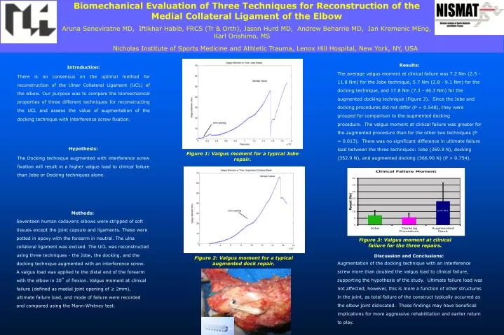
Biomechanical Evaluation of Three Techniques for Reconstruction of the Medial Collateral Ligament of the Elbow Aruna Seneviratne MD, Iftikhar Habib, FRCS (Tr & Orth), Jason Hurd MD, Andrew Beharrie MD, Ian Kremenic MEng, Karl Orishimo, MS Nicholas Institute of Sports Medicine and Athletic Trauma, Lenox Hill Hospital, New York, NY,USA Figure 1: Valgus moment for a typical Jobe repair. Figure 2: Valgus moment for a typical augmented dock repair. p<0.014 Figure 3: Valgus moment at clinical failure for the three repairs. Results: The average valgus moment at clinical failure was 7.2 Nm (2.5 - 11.8 Nm) for the Jobe technique, 5.7 Nm (2.8 - 9.1 Nm) for the docking technique, and 17.8 Nm (7.3 - 46.3 Nm) for the augmented docking technique (Figure 3). Since the Jobe and docking procedures did not differ (P = 0.548), they were grouped for comparison to the augmented docking procedure. The valgus moment at clinical failure was greater for the augmented procedure than for the other two techniques (P = 0.013). There was no significant difference in ultimate failure load between the three techniques: Jobe (369.8 N), docking (352.9 N), and augmented docking (366.90 N) (P > 0.754). Introduction: There is no consensus on the optimal method for reconstruction of the Ulnar Collateral Ligament (UCL) of the elbow. Our purpose was to compare the biomechanical properties of three different techniques for reconstructing the UCL and assess the value of augmentation of the docking technique with interference screw fixation. Hypothesis: The Docking technique augmented with interference screw fixation will result in a higher valgus load to clinical failure than Jobe or Docking techniques alone. Methods: Seventeen human cadaveric elbows were stripped of soft tissues except the joint capsule and ligaments. These were potted in epoxy with the forearm in neutral. The ulna collateral ligament was excised. The UCL was reconstructed using three techniques - the Jobe, the docking, and the docking technique augmented with an interference screw. A valgus load was applied to the distal end of the forearm with the elbow in 30˚ of flexion. Valgus moment at clinical failure (defined as medial joint opening of ≥ 2mm), ultimate failure load, and mode of failure were recorded and compared using the Mann-Whitney test. Discussion and Conclusions: Augmentation of the docking technique with an interference screw more than doubled the valgus load to clinical failure, supporting the hypothesis of the study. Ultimate failure load was not affected; however, this is more a function of other structures in the joint, as total failure of the construct typically occurred as the elbow joint dislocated. These findings may have beneficial implications for more aggressive rehabilitation and earlier return to play.