Download

1 / 34
380 likes | 706 Views
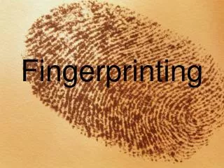
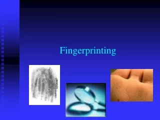
Protein Fingerprinting and Evolution. Ms. Haut. Protein Fingerprinting. Analyze protein profiles from a variety of fish Use acrylamide electrophoresis to separate proteins by size Compare biochemical and phylogenetic relationships. Making Proteins. DNA TAC GGA TCG AGA TGA

E N D
Protein Fingerprinting and Evolution Ms. Haut
Protein Fingerprinting • Analyze protein profiles from a variety of fish • Use acrylamide electrophoresis to separate proteins by size • Compare biochemical and phylogenetic relationships
Making Proteins DNA TAC GGA TCG AGA TGA mRNA AUG CCU AGC UCU ACU tRNA UAC GGA UCG AGA UGA Amino Acid Tyr Gly Ser Arg STOP
1o 2o 3o 4o Levels of Protein Organization
Protein Size Comparison • Break protein complexes into individual proteins • Denature proteins using detergent and heat • Separate proteins based on size
Protein Size • Size measured in kilodaltons (kDa) • Dalton = mass of hydrogen molecule = 1.66 x 10⁻²⁴ gram • Average amino acid = 110 daltons
Actin and Myosin • Actin • 5% of total protein • 20% of vertebrate muscle mass • 375 amino acids = 42 kDa • Forms filaments • Myosin • Tetramer • two heavy subunits (220 kDa) • two light subunits (20 kDa) • Breaks down ATP for muscle contraction Campbell, 2007.
Protein Fingerprinting • First carried out in 1956 to show that sickle-cell anemia was caused by a change in a single amino acid of the hemoglobin protein • The method we will use SDS-PAGE was developed in 1970 by U.K. Laemmli
Protein Electrophoresis http://departments.oxy.edu/biology/Franck/Bio222/Lectures/Feb1lecture.htm • Proteins are charged molecules whose sequences are determined by DNA • Polyacrylamide gels are used to separate small molecules such as proteins • Agarose gels separate large molecules • The percentage of acrylamide gel to be used depends on the size range of the proteins of interest. (the higher the % = the denser the matrix and better the separation of small molecules)
Protein Electrophoresis • Proteins are separated by size—smaller proteins travel farther in the matrix
Useful Information Gathered from Protein Sequence • Relationship to other proteins (protein families) • Example: Viral protein that produces cancer is nearly identical to normal cellular growth factor. • Evolution of organisms (phylogenetic trees). • Information for creating antibodies: specific regions can be identified. • Information for making DNA probes.
Classification Kingdom Phylum Class Order Family Genus Species Traditional classification based upon traits: Morphological Behavioral Traditional Systematics and Taxonomy
Can biomolecular evidence be used to determine evolutionary relationships?
Biochemical Similarities • Traits are the result of: • Structure • Function • Proteins determine structure and function • DNA codes for proteins that confer traits
Biochemical Differences • Changes in DNA leads to proteins with: • Different functions • Novel traits • Positive, negative or no effects • Genetic diversity provides pool for natural selection = evolution
Evolution and Classification of Fishes • Evolutionary trees • Show evolutionary lineages of different species over time http://bio.winona.edu/berg/ILLUST/evoltree.gif
Protein gel electrophoresis • Protein gel from protein fingerprinting experiment. Each blue band represents a distinct protein. The pattern of bands gives information about the composition of a sample http://www.bridgewater.edu/~sbaron/Bio%20325%20Pics.htm
Variations in amino acid sequence in one part of the cytochrome c molecule
Variations in amino acid sequence in one part of the cytochrome c molecule http://www.rtis.com/nat/user/elsberry/evobio/evc/argresp/sequence.html
Fish Classification • Chondrichthyes (cartilaginous fishes) • Cartilaginous skeleton, thick skin w/o scales, no swim bladders or lungs • Sharks, skates, rays • Osteichthyes (bony fishes) • Bony skeleton, true scales, paired fins w/ moveable rays • Agnatha (jawless fishes) • Eel-like, jawless fishes with parasitic and scavenging lifestyles • No scales or paired fins
Phylogenetic tree for fishes http://www.fao.org/documents/show_cdr.asp?url_file=/DOCREP/V7180E/v7180e04.htm
Which fishes should you select? • Choose fish specimens that will provide striking and distinct results • Select some closely related fishes (salmon/trout) and some more distantly related fishes (get exotic—shark, tuna) • Other aquatic organisms—mollusks—scallops, octopus; arthropods—clams, oysters, crab, shrimp
Day 2 Protein Fingerprinting Procedures Day 1 Day 3
What’s in the Sample Buffer? • Tris buffer to provide appropriate pH • SDS (Sodium Dodecyl Sulfate) detergent to dissolve proteins and give them a negative charge • Glycerol to make samples sink into wells • Bromophenol Blue dye to visualize samples http://departments.oxy.edu/biology/Franck/Bio222/Lectures/Feb1lecture.htm
SDS, heat s-s Proteins with SDS – + Why Heat the Samples? • Heating the samples denatures protein complexes, allowing the separation of individual proteins by size
SDS, heat s-s Proteins with SDS – How Does an SDS-PAGE Gel Work? • Negatively charged proteins move to positive electrode • Smaller proteins move faster • Proteins separate by size +
CH3 CH2 CH2 CH2 CH2 CH2 CH2 CH2 CH2 CH2 CH2 CH2 O S O O SDS - O SDS-Polyacrylamide Gel Electrophoresis (SDS-PAGE) • SDS Detergent (Sodium Dodecyl Sulfate) • Solubilizes and denatures proteins • Adds negative charge to proteins • Heat denatures proteins
Why Use Acrylamide Gels to Separate Proteins? • Acrylamide gel give a tight matrix • Ideal for protein separation • Smaller pore size than agarose • Proteins much smaller than DNA • Average amino acid = 110 Da • Average nucleotide pair = 649 Da • 1 kilobase of DNA = 650 kDa • 1 kilobase of DNA encodes 333 amino acids = 36 kDa
Gel Analysis Lane • Kaleidoscope Markers • Shark • Salmon • Trout • Catfish • Sturgeon • Actin and Myosin Standard
kDa mm 203 8.5 135 12.0 86 18.5 41 28.0 33 34.0 19 41.5 8 44.5 Molecular Weight Estimation 250 200 150 Size in kDa 100 50 0 0 20 40 60 Distance (mm from well)
Ready Gel Assembly Step 1 Step 1 Step 2 Step 3 Step 4