Download
1 / 24
320 likes | 941 Views
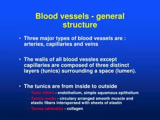
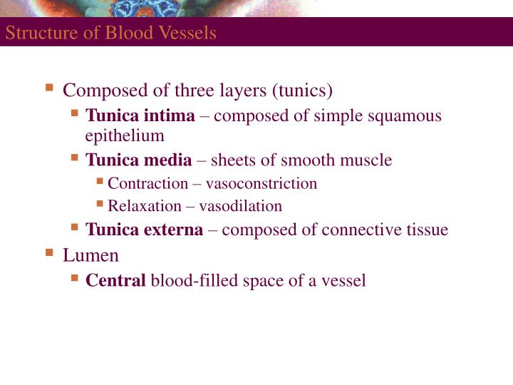
Structure of Blood Vessels. Composed of three layers (tunics) Tunica intima – composed of simple squamous epithelium Tunica media – sheets of smooth muscle Contraction – vasoconstriction Relaxation – vasodilation Tunica externa – composed of connective tissue Lumen

E N D
Structure of Blood Vessels • Composed of three layers (tunics) • Tunica intima – composed of simple squamous epithelium • Tunica media – sheets of smooth muscle • Contraction – vasoconstriction • Relaxation – vasodilation • Tunica externa – composed of connective tissue • Lumen • Central blood-filled space of a vessel
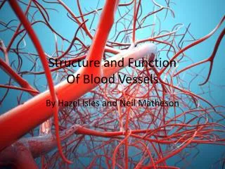
Structure of Arteries, Veins, and Capillaries Figure 19.1a
Types of Blood Vessels • Arteries – carry blood away from the heart • Capillaries – smallest blood vessels • The site of exchange of molecules between blood and tissue fluid • Veins – carry blood toward the heart
Types of Arteries • Elastic arteries – the largest arteries • Diameters range from 2.5 cm to 1 cm • Includes the aorta and its major branches • Sometimes called conducting arteries • High elastin content dampens surge of blood pressure Figure 19.2a
Types of Arteries • Muscular (distributing) arteries • Lie distal to elastic arteries • Diameters range from 1 cm to 0.3 mm • Includes most named arteries • Tunica media is thick • Unique features • Internal and external elastic laminae Figure 19.2b
Types of Arteries • Arterioles • Smallest arteries • Diameters range from 0.3 mm to 10 µm • Larger arterioles possess all three tunics • Diameter of arterioles controlled by • Local factors in the tissues • Sympathetic nervous system Figure 19.2c
Capillaries • Smallest blood vessels • Diameter from 8–10 µm • Red blood cells pass through single file • Site-specific functions of capillaries • Lungs – oxygen enters blood, carbon dioxide leaves • Small intestines – receive digested nutrients • Endocrine glands – pick up hormones • Kidneys – removal of nitrogenous wastes
RBCs in a Capillary Figure 19.3
Capillary Beds • Network of capillaries running through tissues • Precapillary sphincters • Regulate the flow of blood to tissues • Tendons and ligaments – poorly vascularized • Epithelia and cartilage – avascular • Receive nutrients from nearby CT
Capillary Beds Figure 19.4a
Capillary Beds Figure 19.4b
Capillary Permeabillity • Endothelial cells – held together by tight junctions and desmosomes • Intercellular clefts – gaps of unjoined membrane • Small molecules can enter and exit • Two types of capillary • Continuous – most common • Fenestrated – have pores
Structure of Capillaries – Cross Section Figure 19.5a
Structure of Capillaries – Cross Section Figure 19.5b
Routes of Capillary Permeability • Four routes into and out of capillaries • Direct diffusion • Through intercellular clefts • Through cytoplasmic vesicles • Through fenestrations
Low Permeability Capillaries • Blood-brain barrier • Capillaries have complete tight junctions • No intercellular clefts are present • Vital molecules pass through • Highly selective transport mechanisms • Not a barrier against • Oxygen, carbon dioxide, and some anesthetics
Sinusoids • Wide, leaky capillaries found in some organs • Usually fenestrated • Intercellular clefts are wide open • Occur in bone marrow and spleen • Sinusoids have a large diameter and twisted course
Sinusoids Figure 19.5c
Veins • Conduct blood from capillaries toward the heart • Blood pressure is much lower than in arteries • Smallest veins – called venules • Diameters from 8 – 100 µm • Smallest venules – called postcapillary venules • Venules join to form veins • Tunica externa is the thickest tunic in veins
Mechanisms to Counteract Low Venous Pressure • Valvesin some veins • Particularly in limbs • Skeletal muscle pump • Muscles press against thin-walled veins Figure 19.6
Vascular Anastomoses • Vessels interconnect to form vascular anastomoses • Organs receive blood from more than one arterial source • Neighboring arteries form arterial anastomoses • Provide collateral channels • Veins anastomose more frequently than arteries
Vasa Vasorum • Tunica externa of large vessels have • Tiny arteries, capillaries, and veins • Vasa vasorum vessels of vessels • Nourish outer region of large vessels • Inner half of large vessels receive nutrients from luminal blood
Pulmonary Circulation • Pulmonary trunk leaves the right ventricle • Divides into right and left pulmonary arteries • Superior and inferior pulmonary veins • Carry oxygenated blood into the left atrium