Download
1 / 24
260 likes | 513 Views
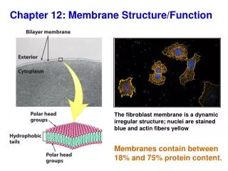
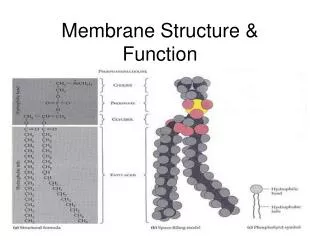
Chapter 8: Membrane Structure and Function. Objectives The student is responsible for: The definitions of all bold faced words in the chapter Knowing the entire chapter. A phospholipid is amphipathic. Figure 8.1 Artificial membranes (cross sections). Outside the cell.

E N D
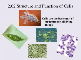
Chapter 8: Membrane Structure and Function • Objectives • The student is responsible for: • The definitions of all bold faced words in the chapter • Knowing the entire chapter.
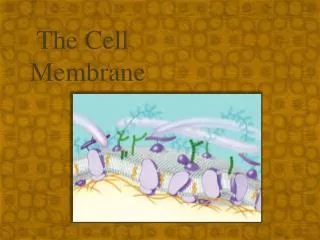
A phospholipid is amphipathic. Figure 8.1 Artificial membranes (cross sections) Outside the cell Phospholipids will naturally form this lipid bilayer thus creating a thermodynam- ically stable boundary. Inside the cell
Figure 8.2 Two generations of membrane models (1935 – 1970’s)
Freeze Fracture allowed researchers to view the split membrane with proteins interspersed within the lipids. Figure 8.3 Freeze-fracture and freeze-etch
Figure 8.4 The fluidity of membranes Membranes are fluid!!!!
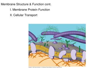
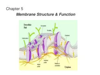
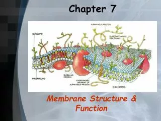
Figure 8.6 The detailed structure of an animal cell’s plasma membrane, in cross section
Structure of a transmembrane protein: alpha helix in the hydrophobic portion Figure 8.7 The structure of a transmembrane protein
Figure 8.8 Sidedness of the plasma membrane Sidedness of the Plasma Membrane 1) external face is similar to inside of the golgi, ER and vesicles 2) CHOs that are made in ER, modified by the golgi end up on external surface.
Membrane CHOs are important for cell-cell recognition • Similar cell types (tissues) need to locate together and therefore they need to recognize each other as they are formed. • Foreign cells need to be rejected. • Small number of sugars are involved (15) • These can be glycolipids or glycoproteins • These molecules help to distinguish tissue from tissue in the same individual, same tissues between individuals and even from species to species. • Blood groups of A, B, AB and O are due to these sugar differences.
Functions of Membrane-Associated Proteins Figure 8.9 Some functions of membrane proteins P. Membrane, organelle’s membrane Chloroplasts or mitochondrial Plasma membrane
Traffic Across Membranes Is Selective • Selective Permeability applies to gases such as oxygen and carbon dioxide and ions, sugars, amino acids, basically all solutes. These substances also move through the membrane at different rates. • Permeability of the Lipid Bilayer • Hydrophobic vs. Hydrophilic • Transport Proteins • these allow for polar molecules and ions to avoid the hydrophobic core • the concentration gradient of that particular substance determines its direction of flow. Each solute follows its own gradient even though the total solute concentration may appear otherwise (fig. 8.10) • Passive transport does not require energy
Bacteria possess membrane pores to take up fatty acids from their environment. The uptake of fatty acids is important for membrane structure, energy and as signaling molecules. • Yes, fatty acids still move across spontaneously. • It is unclear on the these transporters work for the fatty acids. • They do know it has a hydrophobic fatty-acid-binding pocket near the extracellular entrance. No channel connects this pocket to the inside of the cell; instead a hatch plugs the barrel-shaped protein that spans the membrane. It is hypothesized that the hatch changes shape to open up a passageway for the fatty acids to diffuses into the cell. • Source: Chemical and Engineering News, June 7, 2004, pg 30: http://www.CEN-ONLINE.ORG • Researchers: HHMI researcher Tom Rapoport and postdoc Bert van den Berg of Harvard Medical School that crystallized this transporter. • Science, 304, 1506 (2004)
Osmosis Figure 8.11 Osmosis Direction of water flow is determined by total solute concentration, from hypotonic to hypertonic solution
Osmoregulation in Plant and Animal Cells Figure 8.12 The water balance of living cells
Osmoregulation in Paramecium Figure 8.13 The contractile vacuole of Paramecium: an evolutionary adaptation for osmoregulation Filled or filling vacuole Empty vacuole
Facilitated Diffusion Figure 8.14 Two models for facilitated diffusion Some transporters change shape Channel Protein FD requires a transport protein but no energy It is also specific for the solute it is transporting. Aquaporins: channels specific for water; Gated channels require a chemical or electrical stimulus.
Uh Oh!!!!: The sodium-potassium pump: (Will this hurt?) Figure 8.15 The sodium-potassium pump: a specific case of active transport Movie
A summary Figure 8.16 Review: passive and active transport compared
Figure 8.17 An electrogenic pump Or in mitochondria and chloroplasts
Transport of Large Molecules Figure 8.19 The three types of endocytosis in animal cells
Receptor-Mediated Endocytosis • Protein receptors are stuck in the membrane waiting to bind to a specific “ligand” or extracellular substance. • The receptors are in specific regions called coated pits. • Once the ligands bind to the receptors, the coated pit buds off into the cytoplasm, enters the cell and then digested. • Advantage: take in large quantities of a solute even though it is not in high concentration in the extracellular environment. • Example (a very important one) • Cholesterol gets bound to two kinds of molecules in the blood stream: low-density lipoproteins (LDLs) or high-density lipoproteins (HDLs) • Cholesterol bound LDLs bind to LDL receptors on liver cells; get transported into the cell and therefore removed from bloodstream. • Hypercholesterolemia: LDL receptors are defective