Download
1 / 59
610 likes | 929 Views
Membrane Function, Structure & Transport. Cell Membrane (Plasma Membrane). Fluid Mosaic Model of the cell membrane. http://www.susanahalpine.com/anim/Life/memb.htm. About Cell Membranes (continued). Cell membranes have pores (holes) in it

E N D
Membrane Function, Structure & Transport
Fluid Mosaic Modelof the cell membrane http://www.susanahalpine.com/anim/Life/memb.htm
About Cell Membranes (continued) • Cell membranes have pores (holes) in it • Selectively permeable: Allows some molecules in and keeps other molecules out • The structure helps it be selective http://phschool.com/science/biology_place/biocoach/biomembrane1/permeability.html
Structure of the Cell Membrane Outside of cell Lipid Bilayer Inside of cell (cytoplasm)
Membrane Function • Six major functions of membrane proteins: • Transport • Enzymatic activity • Signal transduction • Cell-cell recognition • Intercellular joining • Attachment to the cytoskeleton and extracellular matrix (ECM)
b.Enzymatic activity a. Transport c. Signal transduction
f. Attachment to the cytoskeleton and extra- cellular matrix (ECM) e. Intercellular joining d. Cell-cell recognition
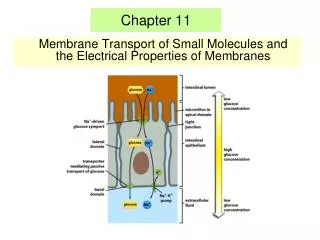
Transport Proteins • Cell membranes are permeable to specific ions and polar molecules, including water. These hydrophilic substances avoid contact with the lipid bilayer by passing through transport proteins that span the membrane • Some transport proteins function by having a hydrophilic channel that certain molecules or atomic ions use as a tunnel through the membrane. • Other transport proteins hold onto their passengers and physically move them across the membrane. • In both cases, the transport protein is specific for the substances it translocates (moves), allowing only a certain substance or class of closely related substances to cross the membrane. • For example, glucose carried in blood to the human liver enters liver cells rapidly through specific transport proteins in the plasma membrane. The protein is so selective that it even rejects fructose, a structural isomer of glucose. • The selective permeability of a membrane depends on both the discriminating barrier of the lipid bilayer and the specific transport proteins built into the membrane.
Proteins • Integral proteins penetrate the hydrophobic core of the lipid bilayer. • Many are transmembrane proteins, which completely span the membrane. The hydrophobic regions of an integral protein consist of one or more stretches of nonpolar amino acids usually coiled into a helices • The hydrophilic parts of the molecule are exposed to the aqueous solutions on either side of the membrane. • Peripheral proteins are not embedded in the lipid bilayer at all; they are appendages loosely bound to the surface of the membrane, often to the exposed parts of integral proteins
Membrane carbohydrates are important for cell-cell recognition • Cell-cell recognition, a cell’s ability to distinguish one type of neighboring cell from another, is crucial to the functioning of an organism. It is important, for example, in the sorting of cells into tissues and organs in an animal embryo. It is also the basis for the rejection of foreign cells (including those of transplanted organs) by the immune system, an important line of defense in vertebrate animals. The way cells recognize other cells is by keying on surface molecules, often carbohydrates, on the plasma membrane. • Membrane carbohydrates are usually branched oligosaccharides with fewer than 15 sugar units. (Oligo is Greek for "few"; an oligosaccharide is a short polysaccharide.) Some of these oligosaccharides are covalently bonded to lipids, forming molecules called glycolipids. (Recall that glyco refers to the presence of carbohydrate.) Most, however, are covalently bonded to proteins, which are thereby glycoproteins.
Membrane carbohydrates are important for cell-cell recognition • The oligosaccharides on the external side of the plasma membrane vary from species to species, among individuals of the same species, and even from one cell type to another in a single individual. The diversity of the molecules and their location on the cellユs surface enable oligosaccharides to function as markers that distinguish one cell from another. For example, the four human blood groups designated A, B, AB, and O reflect variation in the oligosaccharides on the surface of red blood cells.
Diffusion and Osmosis Def:Diffusion - movement of solute from an area of higher concentration to a area of lower concentration http://highered.mcgraw-hill.com/sites/0072495855/student_view0/chapter2/animation__how_diffusion_works.html http://www.indiana.edu/~phys215/lecture/lecnotes/lecgraphics/diffusion.gif
0.55M glucose 0.55M glucose Iain McKillop, Ph.D. imckillo@email.uncc.edu Biol 3111 Diffusion Through Membranes 1. Freely Permeable 0.1M glucose 1M glucose
+ 1M Cl- 0.1M Cl- <1M K+ >0.1M K+ Iain McKillop, Ph.D. imckillo@email.uncc.edu Biol 3111 Diffusion Through Membranes 2. Cation Permeable 1M KCl K+ Cl- 0.1M KCl K+ Cl- K+ K+ Water and K+ CAN move; Cl- CANNOT
Osmosis H20 H20 Iain McKillop, Ph.D. imckillo@email.uncc.edu Biol 3111 Def:Movement of water from a high concentration to a low concentration solution http://zoology.okstate.edu/zoo_lrc/biol1114/tutorials/Flash/Osmosis_Animation.htm Requires a water permeable barrier 0.1M glucose 1M glucose <1M glucose >0.1M glucose “Osmotic Pressure”
Common Terms Def:TONICITY - Measure of the concentration of solutes in a solution Def:OSMOLALITY - Measure of osmotic pressure Def:TURGIDITY - Description of “swollen” state of cells Def:HYPERTONIC - Higher concentration of solute ( dissolved substances) and less solvent Def:HYPOTONIC - Higher concentration of solvent (dissolving substances) and less solute Def:ISOTONIC - Same concentration of solute & solvent Def:plasmolysis -loss of water from a plant
Water Balance of Cells Without Walls • If an animal cell is immersed in an environment that is isotonic to the cell, there will be no net movement of water across the plasma membrane. • Water flows across the membrane, but at the same rate in both directions. In an isotonic environment, the volume of an animal cell is stable. • A hypertonic solution to the cell. The cell will lose water to its environment, shrivel, and probably die. This is one reason why an increase in the salinity (saltiness) of a lake can kill the animals there. • A cell in a solution that is hypotonic to the cell, water will enter faster than it leaves, and the cell will swell and lyse (burst) like an overfilled water balloon.
Isotonic Crenation Hypertonic Hypotonic Osmosis in Red Blood Cells • Observe sheep RBCs via a wet mount of the sample • Aliquot one drop the following solutions with a ½ drop of RBC to a slide • 0.9% saline • 10% NaCl • Distilled water
Water Balance of Cells Without Walls Cont,,, • A cell without rigid walls can tolerate neither excessive uptake nor excessive loss of water. • This problem of water balance is automatically solved if such a cell lives in isotonic surroundings. • Seawater is isotonic to many marine invertebrates. • The cells of most terrestrial (land-dwelling) animals are bathed in an extracellular fluid that is isotonic to the cells. • Animals and other organisms without rigid cell walls living in hypertonic or hypotonic environments must have special adaptations for osmoregulation, the control of water balance.
Contractile Vacuole • Animals and other organisms without rigid cell walls have osmotic problems in either a hypertonic or hypotonic environment • To maintain their internal environment, such organisms must have adaptations for osmoregulation, the control of water balance • The protist Paramecium, which is hypertonic to its pond water environment, has a contractile vacuole that acts as a pump • Video: ChlamydomonasVideo: Paramecium Vacuole
Water Balance of Cells with Walls • The cells of plants, prokaryotes, fungi, and some protists have walls. When such a cell is in a hypotonic solution the wall helps maintain the cell’s water balance. • However, the elastic wall will expand only so much before it exerts a back pressure on the cell that opposes further water uptake. At this point, the cell is turgid (very firm), which is the healthy state for most plant cells. • Plants that are not woody, such as most house plants, depend for mechanical support on cells kept turgid by a surrounding hypotonic solution. If a plant’s cells and their surroundings are isotonic, there is no net tendency for water to enter, and the cells become flaccid (limp), causing the plant to wilt.
Water Balance of Cells with Walls • A wall is of no advantage if the cell is immersed in a hypertonic environment. A plant cell, like an animal cell, will lose water to its surroundings and shrink. As the plant cell shrivels, its plasma membrane pulls away from the wall, (plasmolysis), is usually lethal. The walled cells of bacteria and fungi also plasmolyze in hypertonic environments. Video: Plasmolysis Video: Turgid Elodea Animation: Osmosis
Iain McKillop, Ph.D. imckillo@email.uncc.edu Biol 3111 Membrane Transport 1 Simple Diffusion 2 Facilitated Diffusion 3 Active Transport
Iain McKillop, Ph.D. imckillo@email.uncc.edu Biol 3111 Membrane Transport Simple Diffusion • 1. Small, nonpolar e.g. Oxygen, ethanol • 2. Small, polar • e.g. H2O
Iain McKillop, Ph.D. imckillo@email.uncc.edu Biol 3111 Membrane Transport Facilitated Diffusion *Utilize passive carriers - e.g. channels Ion channels Water channels *Specific binding site for molecules • *Two thermodynamically equivalent shapes • one facing outside, one facing inside *Conformation not affected by solute
Iain McKillop, Ph.D. imckillo@email.uncc.edu Biol 3111 Model of Facilitated Diffusion
Iain McKillop, Ph.D. imckillo@email.uncc.edu Biol 3111 Ion Channels General Properties Selective Fast Passive Gated - i.e. Open or Closed
Facilitated Diffusion of Ions • The transmembrane channels that permit facilitated diffusion can be opened or closed. They are said to be "gated". • Some types of gated ion channels:・ligand-gated・ • Mechanically-gated - examples: • Sound waves bending the cilia-like projections on the hair cells of the inner ear open up ion channels leading to the creation of nerve impulses that the brain interprets as sound • ・Mechanical deformation of the cells of stretch receptors opens ion channels leading to the creation of nerve impulses. • Voltage-gated- In so-called “ excitable cells” like neurons and muscles cells, some channels open or close in response to changes in the charge (measured in volts) across the plasma membrane.Example: As an impulse passes down a neuron, the reduction in the voltage opens sodium channels in the adjacent portion of the membrane. This allows the influx of Na+ into the neuron and thus the continuation of the nerve impulse. Some 7000 sodium ions pass through each channel during the brief period (about 1 millisecond) that it remains open. This was learned by use of the patch clamp technique. • Light-gated- These are triggered by the presence of light. Phototaxis in plants is a result of this type of diffusion.
Iain McKillop, Ph.D. imckillo@email.uncc.edu Biol 3111 Types of Gated Ion Channels
Ligand-gated ion channels • Many ion channels open or close in response to binding a small signaling molecule or "ligand". • Some ion channels are gated by extracellular ligands; some by intracellular ligands. In both cases, the ligand is not the substance that is transported when the channel opens. • External ligandsExternal ligands (shown here in green) bind to a site on the extracellular side of the channel. • Examples: • Acetycholine (ACh). • The binding of the neurotransmetter acetylcholine at certain synapses opens channels that admit Na+ and initiate a nerve impulse or musclecontraction. • ・Gamma amino butyric acid (GABA). Binding of GABA at certain synapses - designated GABAA - in the central nervous system admits Cl- ions into the cell and inhibits the creation of a nerve impulse.
Internal ligands • Internal ligands bind to a site on the channel protein exposed to the cytosol. • Examples: • "Second messengers", like cyclic AMP (cAMP) and cyclic GMP (cGMP), regulate channels involved in the initiation of impulses in neurons responding to odors and light respectively. • ATP is needed to open the channel that allows chloride (Cl-) and bicarbonate (HCO3-) ions out of the cell. This channel is defective in patients with cystic fibrosis. Although the energy liberated by the hydrolysis of ATP is needed to open the channel, this is not an example of active transport; the ions diffuse through the open channel following their concentration gradient.
Iain McKillop, Ph.D. imckillo@email.uncc.edu Biol 3111 Ach Receptor Ligand-gated
Iain McKillop, Ph.D. imckillo@email.uncc.edu Biol 3111 Auditory Hair Cells Stress-gated
H H - - O O K+ H H O O Iain McKillop, Ph.D. imckillo@email.uncc.edu Biol 3111 Channel Specificity
H H - - O O K+ H H O O Iain McKillop, Ph.D. imckillo@email.uncc.edu Biol 3111 Channel Specificity
H H - - O O K+ H H O O Iain McKillop, Ph.D. imckillo@email.uncc.edu Biol 3111 Channel Specificity
H H H H - - - - O O O O H H H H K+ Iain McKillop, Ph.D. imckillo@email.uncc.edu Biol 3111 O O
H H - - O O H H Na+ O O Iain McKillop, Ph.D. imckillo@email.uncc.edu Biol 3111 Channel Specificity
H H - - O O H H Na+ O O Iain McKillop, Ph.D. imckillo@email.uncc.edu Biol 3111 Channel Specificity
H H - - O O H H Na+ O O Iain McKillop, Ph.D. imckillo@email.uncc.edu Biol 3111 Channel Specificity
Ion [Intracellular] [Extracellular] Na+ 5-15 mM 145 mM K+ 140 mM 5 mM H+ 7x10⁵ mM 4x10⁵ mM Mg2+ 0.5 mM 1-2 mM Ca2+ 10-7 mM 1-2 mM Cl- 5-15 mM 5-15 mM Fixed Anions High Low Iain McKillop, Ph.D. imckillo@email.uncc.edu Biol 3111 Chemical Composition of Cells Intra and extracellular compositions are different Very important in removing waste and letting required molecules in Unequal distribution of ions is critical to cell function
Iain McKillop, Ph.D. imckillo@email.uncc.edu Biol 3111 Active Transport *Active transport moves molecules AGAINST a gradient *Essential for cell function - Moves molecules IN and OUT *Requires energy Pumps Coupled Transport
Active Transport 1. Pumps Energy provided by LIGHT or ATP http://www.wisc-online.com/objects/index_tj.asp?objID=AP11203
Iain McKillop, Ph.D. imckillo@email.uncc.edu Biol 3111 Active Transport 1. Pumps Na+/K+ Exchange pump is VERY important Acts as a pump AND an enzyme
The sodium-potassium pump: a specific case of active transport. • This transport system pumps ions against steep concentration gradients. The pump oscillates between two conformational states in a pumping cycle that translocates three Na+ ions out of the cell for every two K+ ions pumped into the cell. ATP powers the changes in conformation by phosphorylating the transport protein (that is, by transferring a phosphate group to the protein).
Active Transport Protein Pumps-transport proteins that require energy to do work • Example: Sodium / Potassium Pumps are important in nerve responses. Protein changes shape to move molecules: this requires energy!
Some ion pumps generate voltage across membranes • All cells have voltages across their plasma membranes. Voltage is electrical potential energy--a separation of opposite charges. The cytoplasm of a cell is negative in charge compared to the extracellular fluid because of an unequal distribution of anions and cations on opposite sides of the membrane. The voltage across a membrane, called a membrane potential, ranges from about -50 to -200 millivolts. (The minus sign indicates that the inside of the cell is negative compared to the outside.)
Voltage cont.. • The membrane potential acts like a battery, an energy source that affects the traffic of all charged substances across the membrane. • Because the inside of the cell is negative compared to the outside, the membrane potential favors the passive transport of cations into the cell and anions out of the cell. • Two forces drive the diffusion of ions across a membrane: a chemical force (the ion’s concentration gradient) and an electrical force (the effect of the membrane potential on the ion’s movement). • This combination of forces acting on an ion is called the electrochemical gradient. • An ion does not simply diffuse down its concentration gradient, but diffuses down its electrochemical gradient. For example, the concentration of sodium ions (Na+) inside a resting nerve cell is much lower than outside it. When the cell is stimulated, gated channels that facilitate Na+ diffusion open. Sodium ions then "fall" down their electrochemical gradient, driven by the concentration gradient of Na+ and by the attraction of cations to the negative side of the membrane.
Voltage Cont.. • Some membrane proteins that actively transport ions contribute to the membrane potential. • An example is the sodium-potassium pump. • The pump does not translocate Na+ and K+ one for one, but actually pumps three sodium ions out of the cell for every two potassium ions it pumps into the cell. With each crank of the pump, there is a net transfer of one positive charge from the cytoplasm to the extracellular fluid, a process that stores energy in the form of voltage. • A transport protein that generates voltage across a membrane is called an electrogenic pump. The sodium-potassium pump seems to be the major electrogenic pump of animal cells. The main electrogenic pump of plants, bacteria, and fungi is a proton pump, which actively transports hydrogen ions (protons) out of the cell. The pumping of H+ transfers positive charge from the cytoplasm to the extracellular solution • http://www.wiley.com/legacy/college/boyer/0470003790/animations/membrane_transport/membrane_transport.htm