Download
1 / 28
280 likes | 364 Views
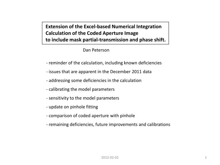
Extension of the Excel-based Numerical Integration Calculation of the Coded Aperture Image to include mask partial-transmission and phase shift. Dan Peterson. - reminder of the calculation, including known deficiencies - issues that are apparent in the December 2011 data

E N D
Extension of the Excel-based Numerical Integration Calculation of the Coded Aperture Image to include mask partial-transmission and phase shift. Dan Peterson - reminder of the calculation, including known deficiencies - issues that are apparent in the December 2011 data - addressing some deficiencies in the calculation - calibrating the model parameters - sensitivity to the model parameters - update on pinhole fitting - comparison of coded aperture with pinhole - remaining deficiencies, future improvements and calibrations 2012-02-02 1
History: calculated image Recall, the image calculation and fitting technique were developed in January 2011, uses an Excel spreadsheet to perform the numerical integration of the amplitudes, (1) where y is the vertical position at the plane of the optics element, r(y) is the path length from the source, through the optic, to the detector pixel, T(y) is the transmission of the optics element as a function of vertical position. E = E0 ∑ e i 2π r(y) /λT(y) Pulse height is calculated for 64 pixels of size 25μm, (half diodes) . Paths are separated by Δy=0.5 μm at the optic. (Features are 10 μm. ) January 2011 version had many shortcuts: x-ray energy: mono-energetic 2.0 keV, λ=6.2 x10⁻¹⁰ m, 6.2 x10⁻⁴ microns. transmission of the gold coded aperture mask is 0 the semitransparent mask contributes to the diffraction pattern range of the integration is ±155 microns, projects to ± 0.52 mm at the detector. The integration produces the point source image at right. raw smoothed 50% n.n. 2012-02-02
History: Sum Of Gaussian fit The beam size=0 image is parameterized as a Sum Of Gaussians, SOG. The parameterization allows a simple convolution with the broadening due to beam size. The fitting function is then: F = B + C / (2π)½∑J=1,12 AJ / (σJ2 + σ2)½ e -½ ( x - X0J - X0)2 / (σJ2 + σ2 ) (2) The 36 function definition parameters are calculated, for beam size=0, once. These parameters are ( AJ , σJ , X0J ) J=1,12 . In January 2011, there were 4 fitting parameters: C (area), X0 (position), σ (magnified beam size), B (background ) 2012-02-02 3
History: Recalibrating (fudging) the calculated image The original calculated image had systematic deviations from the data. We expected inaccuracies in the calculated image from the shortcuts: use of mono-energy and opaque mask, The side bumps, in particular, would be enhanced with the inclusion of semi-transmission. An empirical “recalibration” was applied to improve the fit. December 2010 D-line, 14ns readout image, as calculated image, after recalibration Although stepping away from a calculation with known inputs, the recalibration provides a point-source image closer to reality. This reduces the minimum χ2 and makes the fit more sensitive to the beam size. 2012-02-02 4
Performance in January 2011 Results from the 3 optics elements were similar. 2012-02-02 5
Attempts to fit December 2011 data. (3) interference dip - is lower than the fit, is lower than the background level uncover several problems • primary peak is OK, • but is dominating the fit (4) left shoulder of the primary peak - is higher and narrower than the fit (2) secondary peak - is higher than the fit (5) background - does not match a flat line - is from a material property and should be a fixed contribution 004018, BigD Shown: 3 conditions, 4 turns each 004020, BigD displaced detector 004029, Norm 2012-02-02 6
Improving the image calculation in January 2012. The image shape should be calculated from characteristics of the x-ray beam and the coded aperture, without an ad-hoc recalibration. Add an x-ray energy distribution and the coded aperture mask semi-transmission. The range of integration at the plane of the mask was expanded to project beyond detector boundaries. The background is a fundamental problem with the original fit. The image fills the detector so there is not an independent measure of the background in a single pass. The floating background allows degenerate solutions; the two main peaks and the dip between them can be fit with a smaller width and larger background. It would be nice to be able to apply everything we know about the beam, filters/windows and the mask, and get the right answer. But, for example, the mask thickness specification appears to be inaccurate. The semi-transmission of the mask should be calibrated to the data. Image shape details may depend on the personality of a specific coded aperture. The semi-transmission is accompanied by a phase shift of waves passing through the mask. As will be described, the phase shift is significant, and significantly changes the image shape. For example, the dip between the two main peaks is enhanced by interference. (Someone thought about this when designing the coded aperture.) The phase shift provides an extra handle in calibrating the semi-transmission of the mask. 2012-02-02 7
Calibrating the image function I take an empirical, data driven , approach to calibrating the image function. Continue using Excel, which provides rapid feedback. The limitation of Excel is that the energy spectrum is limited to 5 discrete energies. calibration variables: For the current calibration, these variables are tuned without much theoretical input. (1) low energy cut-off of the x-ray spectrum (2) high energy tail of the x-ray spectrum (3) semi-transmission of the coded aperture mask (4) phase shift in passing through the gold mask 004029, Norm, turn 1 There are still shortcuts: The transmission and phase shift are ( as yet ) energy-independent. The goal of the calibration is to match major features of the observed image (1) primary peak (2) secondary peak (3) interference dip (4) left shoulder of the primary peak (5) background (6) side peak at channel 7 (7) side peak at channel 31 2012-02-02 2012-02-02 8 8
You may ask, “Why not just use John’s image?” I have parameterized John’s image with 12 gaussians. Some of the smaller fine features (< 1 pixel) are smoothed out. The image is available as an alternative function in CodedAperatureFit.m 2012-02-02 9
You may ask again, “ So why not just use John’s image?” The fit to the left shoulder of the primary satellite is improved, compared to the DanP original fit. The fit to the secondary peak is low. The fit at the central dip does not reach low enough. The fit does not account for the background. Note that the fit to the peak is high. (Fit to displaced detector uses beam size constrained to result from centered detector fit.) 004018, BigD 004020, BigD displaced detector The floating background is a source of uncertainty. We can use John’s image with an added, even locked, background. However, there are the shape issues described above. The apparent underestimation of the transmission indicates that the phase is incorrect. (The discussion of phase is coming… next.) 2012-02-02 10
transmission, phase shift, and the complex index of refraction ref. X-ray Data Booklet, LBNL/PUB-490 The expression for the complex index of refraction n = 1 - δ - iβ (3) describes the phase velocity of the wave and absorption due to atomic interactions. The phase velocity is greater than c, speed of light in a vacuum. The wave equation (1) stated in slide 2 E = E0 ∑ e i 2π r(y) /λT(y) (1) becomes (including the time dependence) E = E0 ∑ e i (2π / λ) (-ct + r) e - i (2π / λ ) δ r e – ( 2π / λ ) β r (4) So far, this is simply an expression of the undisturbed wave, a phase shift, and the transmission. 2012-02-02 11
transmission, phase shift, and the complex index of refraction ref. X-ray Data Booklet, LBNL/PUB-490 The transmission and phase shift terms are calculated from atomic scattering of the x-rays and expressed as δ = reλ2 na / (2π) f1 (5) iβ = i reλ2 na / (2π) f2 (6) where re is the electron radius, na is the atomic number density and f1 and f2 are unit-less form factors, which are provided in the reference in graphs. Substituting the definitions, (5) and (6) into equation (4) E = E0 ∑ e i (2π / λ) (-ct + r)e - i 2π (reλ na 1/(2π) f1 ) r e – ( reλ naf2 ) r (7) We can calculate the image shape with this equation, knowing the energy spectrum and material thicknesses (windows and coded aperture) . footnote: Noting the similarity of the phase shift and transmission terms, the wave equation can be expressed in a form that is dependent on a measured amplitude transmission: T = A/A0 = e – ( reλnaf2 ) r(8) then E = E0 ∑e i(2π / λ) (-ct + r(y) ) T(y,λ)e i 2π [ ln( T(y,λ) ) 1/(2π) f1 (λ) / f2 (λ) ] (9) 2012-02-02 12
calculation of transmission and phase shift: what is reasonable for the specification gold thickness footnote: Alternatively, the transmission can be calculated from the macroscopic description: T = ( I / I0 ) ½ = e - ½ μρ r μ is taken from the plot in the reference: 2.8 x 103 cm2/g T (2.4 keV) = e-1.891 = 0.151 Thus, the mass absorption coefficient , μ , is related to the form factor, f2, by μ = 2 re N/Aauλf2 Amplitude transmission and phase shift can be calculated from equation (7): E = E0 ∑ e i (2π / λ) (-ct + r)e - i 2π (reλ na 1/(2π) f1 ) r e – ( reλ naρf2 ) r (7) f2 phase shift transmission The amplitude transmission is thus T = e – ( reλ naf2 )r = e – ( reλ N/AAuρf2 )r where r is the thickness of the gold: specification: 0.7 x 10⁻⁴ cm re is the electron radius: 2.818 x 10⁻¹³ cm N is Avogadro’s number 6.022 x 10+23/g AAu is the atomic weight of gold: 197 ρ is the density of gold, 19.3 g/cm3 f2 is taken from the plot: 31 at 2.4 keV , λ=5.17 x10⁻⁸ cm 10 at 2.0 keV , λ=6.20 x10⁻⁸ cm T (2.4 keV) = e-1.865 = 0.155 andT (2.0 keV)= = 0.49 ( the simple average is 0.32 ) μ (cm2/g) I express the phase shift as a fraction of 2π. Again, referring to equation (7): θ = - ( reλ na 1/(2π) f1 ) r = - ( reλ N/AAu ρ 1/(2π) f1 ) r f1 is taken from the plot: 52 at 2.4 keV , λ=5.17 x10⁻⁸ cm 50 at 2.0 keV , λ=6.20 x10⁻⁸ cm θ (2.4 keV) = -0.50 θ (2.4 keV) = -0.57 2012-02-02 13
Calculation of transmission and phase shift: what is reasonable for the observed background (the gold thickness issue) The specification gold thickness, 0.7 micron, leads to an average transmission that does not account for the large background under the coded aperture image. The data indicates a lower gold thickness, ~0.5 micron, approximating with an energy-independent transmission and phase. (As described earlier, the current calibration uses average values for the transmission and the phase.) for illustration, this actually corresponds to ~0.8 micron gold thickness. The plots at the right, show the transmission and phase shift for 0.5 micron gold thickness. Loosely averaging {1.7 to 4.0 keV} leads to an expected average amplitude transmission of ~0.43 and average phase shift of ~ -0.35 x 2π. 2012-02-02 14
Input Energy distribution: other material in the beamline Other material in the beam line are 2.5 microns of Silicon in the Coded Aperture substrate, ~6 ??? microns of Carbon in the diamond window, and 0.16 microns Si3N4 in the diode passivation layer, which I ignore. I would like to express this in terms of equivalent carbon because I have a spread sheet. Using the mass absorption coefficient, μ , T = ( I / I0 ) ½ = e - ½ μρ r Mass absorption coefficient are shown in the graphs at right. The ratio for Silicon to Carbon is: μSiρSi / μCρC = ( 995 x 2.33) / (105 x 3.52 ) = 6.27 at 2.5 keV ( the slopes are similar ) Ignoring the Silicon K-edge, 2.5 microns of silicon has roughly the absorption equivalent of 16 microns of carbon. μSi μC 2012-02-02 15
Input Energy distribution, what I use The power distribution is calculated (at upper right) with 6 micron and 22 micron carbon filtering. (Aaron Lyndaker) The combined diamond window and silicon is represented by the 22 micron line (ignoring the 1.839 keV Silicon K-edge). (6 micron line represents the diamond window used for the pinhole.) Brian Heltsley is improving the power distribution to include the silicon and gold K-edges. Future calculations should include the more realistic power distribution, and energy dependent gold transmission and phase shift. But, for now… The numerical integration includes 5 discrete energy values (lower right). In the red distribution, the 5 delta functions are spread into equal-area blocks centered on the discrete energies. This is the energy distribution used in the current calculation, along with energy-independent gold amplitude transmission and energy-independent phase shift. 2012-02-02 16
The current coded aperture image and Sum Of Gaussians 2012-01-27 calculate image with photon energy distribution, 1.9, 2.0, 2.2, 2.5, 2.9, (ave=2.30), scaled to average energy=2.40 amplitude transmission of mask = 0.450 phase shift = -0.31 ( x 2π ) “pulse height uncertainty” = 351.48 (this stuff is for my records) 2012-02-02 17
Fits of the resulting function to beam various conditions 004018, BigD ~18 micron The background basically follows the shape of the data, but now may be slightly high and may require wider shape. Features in the very low beam size data mainly fit well, but the dip in the fit may be too low. 004020, BigD displaced detector 004029, Norm ~13 micron 021442 Coup8 ~23 micron 2012-02-02 18
Image contributions from the 5 energies. with 0.45 amplitude transmission, at all energies with -0.31 x 2π phase , at all energies 1.98, 2.09, 2.3, 2.61, 3.03 keV with average= 2.4 keV In each case, the red line is the parameterization of the sum. 1.98 keV 2.09 keV 2.30 keV 2.61 keV 3.03 keV And, the possible contribution from lower and higher energies 1.6 keV 4.0 keV 2012-02-02 19
variation with phase all with 0.45 amplitude transmission, at all energies phase = 0 phase = -0.1 phase = -0.2 phase = -0.3 phase = -0.4 phase = -0.5 phase = -0.8 phase = -0.9 phase = -0.6 phase = -0.7 2012-02-02 20
variation with gold mask thickness 0.2 0.3 0.5 0.7 1.0 micron gold all with standard energy distribution, transmission ~ e-thickness phase ~ thickness In each case, the red line is the parameterization of the standard image, artificially shifted to match the background. Gold thickness ~ 0.2 micron Gold thickness ~ 0.3 micron Gold thickness ~ 0.5 micron Gold thickness ~ 0.55 micron Gold thickness ~ 0.7 micron Gold thickness ~ 1.0 micron 2012-02-02 21
compare pinhole and CA on coup 8 scan pinhole fit: compare with/without added wide part With smaller beam size in the December 2011 running, a non-gaussian component to the diffraction pattern became visible. A 2nd wide gaussian component was added to the fit. This changes the fitted width of the main component. sigmarms back with 20% wide 0.8824 0.879 0.092 without 0.9905 1.034 0.132 The current pinhole subtractor is 16 microns. What would the subtractor be with the 2011 fit: single gaussian with floating background ? (20 x 0.8824)2 -162 = (20 x 9905)2 –S2 S = 18.4 So, the reduction of the subtractor from 21 to 16 is half due to the function change. 2012-02-02
compare pinhole and CA on coupling-8 scan subtractor = 16 microns 2012-02-02 23
summary, future improvements, calibrations The new coded aperture function has only 3 free parameters, no floating background. It provides stable fits up to 55 micron beam size. The main features have sizes of about 8 micron (referred to the source) , which should provide stable beam size measurements to about 6 microns. (The pixel size will be a significant effect below 6 microns.) Even though this is currently using average values, the transmission and phase of the resultant calculated image match the expectations of the averages better than might be expected. The calculated image was “fit” to the data before investigating the range of reasonable values for the amplitude transmission and the phase. We plan to input a detailed energy spectrum seen by the coded aperture, add energy-dependent transmission and phase shift, geared to a variable input gold thickness. Various experiment can be done to understand the transmission and phase: take measurements with various filters, measure the details of the image from the “box” on the optics chip, take offset images (moving the detector) to efficiently place the major peaks and sufficient background. Daily monitoring the image must be performed to test for changes in the transmission. 2012-02-02 24
Calibration with the “box” on the optic chip variation with phase; energy distribution as in current image calculation all with 0.45 amplitude transmission, 0.20 intensity transmission phase = 0 phase = -0.1 phase = -0.2 phase = -0.3 phase = -0.4 phase = -0.5 phase = -0.8 phase = -0.9 phase = -0.6 phase = -0.7 2012-02-02 25
backup 2012-02-02 26
In each case, the blue line is the new image, 2012-01-27. The red line is the SoG of the January 2011 image (narrow and smoothed) Added 1200 background, scaled by 0.9, for the overlay No tweak With tweak (+.5, x2.5) , (+0, x0.5), (+1.5, x1.5) 2012-02-02
2012-01-31 Set up function CodedAperatureFit.m with JohnF2 (gaussian background in parameterization) DanP6 (.45 trans, -.31phase(on each wavelength) ) Both have ZERO added background. DanP6, 2 events each, 4029, 21442 get also 004018 JohnF2, 2 events each, 4029, 21442 put this in backup