Download
1 / 18
180 likes | 592 Views
PATIENT MANAGEMENT FRAMEOWORK CASE PRESENTATION. NAME : Jason Bartley, DPT, OCS, COMT DATE:10/17/12 BODY REGION : shoulder. Case Rationale.

E N D
PATIENT MANAGEMENT FRAMEOWORK CASE PRESENTATION NAME: Jason Bartley, DPT, OCS, COMTDATE:10/17/12BODY REGION: shoulder
Case Rationale • I chose this case to present because it demonstrates how even after subjective exam and physical exam, I adjusted my initial treatment strategy based on patient preference. Interested most in feedback on this decision. Historically, I would have really focused the GH joint on day 1 treatment.
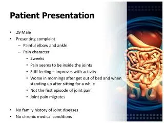
PATIENT PROFILE Patient Profile:Patient is a 17-year old male high-school student. He reports that he is right hand dominant and that prior to this injury he participate in varsity wrestling and football. Patient was referred for left shoulder pain following a closed, non-displaced proximal humerus fracture. Chief complaint: Patient states “The top of my shoulder and my middle/upper back have pain and tightness when I lift or twist my arm”. Date of Injury: 7 weeks ago Self Reported Scores / Outcome Tools: (ODI, NDI, LEFS, DASH, etc) Pt FOTO score = 47 Mean Score = 54 Fear = high Predicted change = 29 Predicted # visits = 13 Predicted length of episode (days) = 41
BODY DIAGRAM • Primary complaint (s) in depth: • P1: intermittent superior shoulder pain with active elevation of the shoulder or external rotation of the shoulder, 0/10 at rest, 7/10 at worst after repetitive lifting (no weight), eases OOP after 3-5 minutes if 7/10 or immediately if only lifting one time • P2: intermittent left scapular “stiffness” at the inferior border, 0/10 at rest, 4/10 at worst with endrange forward elevation of the left shoulder, eases 1-2 minutes OOP • Numbness and or Tingling: denies • Relationship between symptom areas: P1 present as painful arc, while P2 only present at endrange elevation. P1 P2
PE Planning I. What areas/structures must be considered a source of symptoms?
Symptom Behavior • Aggravating and Easing Factors: • P1 Aggravating Factors: • 1: active shoulder elevation • 2: repetitive reaching when doing the dishes at home • 3: performing stretches that were prescribed by MD • 4: rotation/reaching his arm out to the side • P1 Easing Factors: • 1: not using the left arm • P2 Aggravating Factors: • 1: stretching his shoulder all of the way to the end of his range • P2 Easing Factors: • 1: avoiding endrange elevation
History • Sleep and 24 hour pattern: • Sleep: no trouble falling asleep/staying asleep. Normal side sleeper • Morning: no pain upon rising in the morning • Day: depends of activity, specifically overhead elevation • Duration of current symptoms: 1 week • Mechanism of injury / current history: Patient was engaged in wrestling match when he was thrown directly on the lateral side of the left shoulder with his arm wrapped behind his back. Imaging revealed proximal humerus fracture, treated with 7 weeks immobilization, PT not order until after 7 week follow-up.
PATIENT INTAKE • Medical History / Co-Morbidities / Review of Symptoms (ROS): • No signficant medical history, no previous UQ injuries • Red Flag Screen: (-) red flag screen • Yellow Flag Screen: (-) depression screen, though FOTO intake reveales high FEAR score • Special Questions: • Diagnostic tests / Imaging : recent imaging reveals good healing along fracture site, cleared for AROM/strengthening • Medications : NSAIDS PRN
Subjective Asterisks • What will you use as your asterisk signs from the history? (Specify for P1, P2, etc) • P1: superior shoulder pain with active elevation • P1: superior shoulder pain active ER • P2: stiffness at endrange shoulder elevation
Clinical Reasoning • P1/P2 • (S) What is the severity of the condition (min, mod, severe)? • Moderate as it is only present with specific movement, though the pain is keeping him from normal recreational/sport participation • (I) What is the irritability of the condition (min, mod, severe)? • Input your answer and justification: minimal as the pain is only brought on with specific movement and eases very quickly OOP. Pain is present with one repetition and worsens with repeated movement • (N) What is your primary nature statement of the problem - mechanical nocioception • (S) What is the stage of the disorder: sub acute stage • (S) What is the current stability of the disorder • Stable and reproducible with mechanical component
Planning the PE • What will you include to rule in/out your hypotheses? • 1: Impingement TIC • 2: RTC TIC • 3: cervical radic CPR • 4: AC joint special testing • 5: palpation • 6: resisted muscle testing
Physical Exam • Precautions and or Contraindications: healing fracture • Postural Observation: significant thoracic kyphosis, forward head and protracted shoulders • Quick screening tests / clearing of additional joint structures: HBH=P1/P2, HBB=P1 only at mid thoracic, limited compared to R UE. • Neurological Examination:normal dermatome exam, equal/responsive MSR • ROM: • Active left shoulder flexion: 132 degrees *P1/P2 • Active left shoulder ABD: 110 degrees * P1/P2 • Active left shoulder ER: 28 degrees *P1 • Active left shoulder IR: 30 degrees, posterior shoulder stiffness only, unlike P1/P2 • Cervical spine: only mild limitation in flexion with c/o “tightness” around CTJ area, no reproduction of P1/P2. Other planes cleared with sustained OP and quadrant.
Physical Exam • Palpation: (-) AC joint, RTC insertion, left rib angles, (+) TTP along inferomedialscap border (rhomboid, lats) • Spinal Segmental and or Joint Restrictions: Hypomobility noted with CPA at CTJ and upper t-spine, no reproduction. ** Patient very hesitant to allow GH joint mobility testing due to fear of reinjury.** • Manual Muscle testing: * Reproduction of P2 with resisted testing of lats and rhomboid in lengthened position. • Motor control: Displays increased upper trap activation with shoulder elevation • Special tests: (-) Spurlings, (-) ULTT2B (modified for shoulder ROM), (-) cervical distraction, (-) AC joint, (+) Hawkins-Kennedy, (+) ER resistance, (-) drop arm
Assessment & Plan • What is your primary hypothesis following the PE as well as the competing hypothesis (include contributing and predisposing factors as well): • Patient presents with signs consistent with likely secondary shoulder impingement due to poor scapulohumeral kinematics folllowing 7 weeks of immobilization. Also displays secondary impairment in scapular muscle length/strength, kinematics and CT spine mobility, all likely secondary to MOI and immobiliztion • ** Overall patient appears to be healing very well with no c/o TTP or aching/pain near fracture site. However, patient has elevated fear of likelihood that he will reinjure his shoulder if he does too much too soon or if I direct treatment to the “shoulder bone”. Being that patient preference is part of EBP paradigm, along with strong feeling that I can produce quick improvements in asterisk signs with treatment directed away from GH joint, I plan to begin with treatment targeted to the CT spine and scapula.
Day 1 Treatment Treatment provided today and the patient’s response to each intervention. TREATMENTS Rx 1: seated, modified ( L arm at side) CTJ and thoracic distraction) Rx 2: sidelying scapular mobilizations, emphasizing posterior tilt and abduction RESPONSES TO… Rx 1: active shoulder flexion to 138 degrees Rx 2: active shoulder flexion to 150, minimal P2 at end range (2/10).
Response & Assessment at Day 1 • Other treatment provided today and the patient’s response: • Exercise: scapular clocks and retraction for HEP • Education: education of healing phases after fracture, discussed findings of recent radiograph • What changes did you note in your asterisks (test/retest)? • Significant improvement in active shoulder elevation with decreased NPRS • Prognosis (note timeframe of expected level of recovery): • Given history of good healing with fracture and overall age and activity level, I expect very good prognosis. However, I am cautioned by patients concern with fear of reinjury. I got the impression that after the end of day 1 that he was very happy with in session improvement and education seemed to really “get through” and I feel I gained considerable trust/rapport with this patient • Plan of care (including plan for assessment on day 2): • Better: continue CT and scapular manual therapy, begin GH joint mobility, begin active scapulothoracic training/strengthening • Worse: trial CT graded mobilization • Same: continue day 1 treatment, begin and emphasize GH joint mobility