Download
1 / 18
180 likes | 362 Views
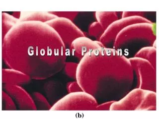
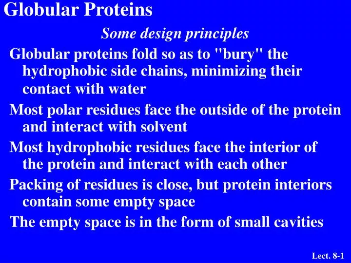
Globular Proteins. Some design principles Globular proteins fold so as to "bury" the hydrophobic side chains, minimizing their contact with water Most polar residues face the outside of the protein and interact with solvent

E N D
Globular Proteins • Some design principles • Globular proteins fold so as to "bury" the hydrophobic side chains, minimizing their contact with water • Most polar residues face the outside of the protein and interact with solvent • Most hydrophobic residues face the interior of the protein and interact with each other • Packing of residues is close, but protein interiors contain some empty space • The empty space is in the form of small cavities
Globular Proteins • More design principles • "Random coil" is not random • Structures of globular proteins are not static • Various elements and domains of protein move to different degrees • Some segments of proteins are very flexible and disordered. • Myoglobin and hemoglobin are typical examples of globular proteins. • Both are heme-containing proteins and each is involved in oxygen metabolism.
Myoglobin: 2o and 3o aspects • Myoglobin is a single peptide chain of 153 residues arranged in eight a-helical regions labeled A-H. • The heme cofactor is the oxygen binding site so it is necessary for myoglobin’s function, oxygen storage in mammalian muscle tissue. • His E7 and F8 are important for binding the heme group within the protein and for stabilizing bound oxygen.
The Heme Group Pyrrole ring - - C H C H C O O O O C C H C H 2 2 2 2 C H H C 3 3 N N Fe(II) H C N N 2 C H C H 3 C H C H C H 3 2
Binding Site for Heme N of His F8 binds to 5th coordination site on heme iron • Oxygen binds to 6th coordination site on heme iron
Oxygen binding to heme His E7 acts as a gate to favor oxygen binding over carbon monoxide.
Hemoglobin • A tetrameric protein • two a-chains (141 AA) • two b-chains (146 AA) • four heme cofactors, one in each chain • The a and b chains are homologous to myoglobin. • Oxygen binds to heme in hemoglobin with same structure as in Mb but cooperatively: as one O2 is bound, it becomes easier for the next to bind.
Hemoglobin: ribbons + hemes • Each chain is in ribbon form. • The heme groups are in space filling form
Oxygen Binding Curves • Hemoglobin and myoglobin respond differently to increase in O2 concentration. • Myoglobin shows normal saturation behavior while hemoglobin shows cooperative behavior. Each oxygen added to a heme of Hb makes addition of the next one easier. • The myoglobin curve is hyperbolic. • The hemoglobin curve is sigmoidal.
Oxygen binding by hemoglobin A Quaternary Structure Change One alpha-beta pair moves relative to the other by 15 degrees upon oxygen binding This large change is caused by movement of Fe by only 0.039 nm when oxygen binds
The Bohr Effect • Competition between oxygen and H+ • Discovered by Christian Bohr • Binding of protons diminishes oxygen binding • Binding of oxygen diminishes proton binding • Important physiological significance-O2 saturation of Hb responds to pH
Bohr Effect II • Carbon dioxide diminishes oxygen binding • CO2 produced in metabolically active tissue (requires oxygen) • Hydration of CO2 in tissues and extremities leads to proton production • CO2 + H2O HCO3- + H+ • These protons are taken up by Hb forcing more oxygen to dissociate • The reverse occurs in the lungs
2,3-Bisphosphoglycerate An Allosteric Effector of Hemoglobin The sigmoid binding curve is only observed in the presence of 2,3-BPG Since 2,3-BPG binds at a site distant from the Fe where oxygen binds, it is called an allosteric effector
2,3-bisphosphoglycerate (2,3-BPG) is a negative allosteric effector of O2 binding to Hb - binds tighter to deoxyHb 2,3-BPG