Download
1 / 45
450 likes | 732 Views
Globular Proteins. What are we going to learn from this chapter?. Myoglobin (Mb) Structure of Heme Structure and Function of Hemoglobin (Hb) Binding of oxygen with Hb and Mb Allosteric Effectors Bohr Effect Variants of Hb Hemoglobinopathies. Types of proteins.

E N D
What are we going to learn from this chapter? Myoglobin (Mb) Structure of Heme Structure and Function of Hemoglobin (Hb) Binding of oxygen with Hb and Mb Allosteric Effectors Bohr Effect Variants of Hb Hemoglobinopathies
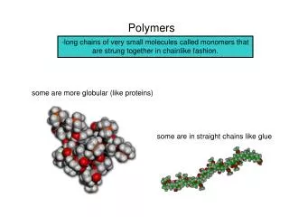
Types of proteins Simple – purely amino acids Complex – with a prosthetic group Globular -has hydrophobic pocket Example: Hemoglobin, Myoglobin Fibrous -repetitive Example: collagen Disease: OI Membrane–spanning -transmembrane (7-helix motif common) Example: insulin receptor Disease: Diabetes
Myoglobin (Mb) Myoglobin is a monomeric heme protein found mainly in muscle tissue where it serves as an intracellular storage site for oxygen. A myoglobin polypeptide is comprised of 8 separate right handed -helices, designated A through H, that are connected by short non helical regions.
Myoglobin (Mb) O2 distal his proximal his Mb is present in both heart and skeletal muscle heme
Heme Heme = protoporphyrin IX + Fe (II)
Mb If heme binds O2, what is the purpose of the peptide/protein portion in Mb? Y partial pressure of oxygen A hyperbolic O2 binding curve indicates a strong affinity towards O2. Even at low oxygen pressure, Mb would sequester the O2 right away making Mb well-suited to its function. [MbO2] Y = [MbO2 + Mb]
A Question on Mb Why do whales need to have a very high concentration of Mb in their muscles?
Hemoglobin (Hb) : A molecule to breath with Hemoglobin is an [(2):(2)] tetrameric hemeprotein found in erythrocytes where it is responsible for binding oxygen in the lung and transporting the bound oxygen throughout the body where it is used in aerobic metabolic pathways.
Hb 2 and 2 subunits 4 heme groups in one Hb molecule 4 O2 molecules can potentially bind to one Hb molecule
and chains of Hb 15126 141 Arg Val-leu 7 Adequate for entry of one molecule of O2 15866 146 His Val-his-leu 8 Entry of O2 in heme pocket is blocked by a val residue Mol weight Total amino acids C-ter residue N-ter residue -helices Heme pocket
Why Mb for storage and Hb for transport? A sigmoidal O2 binding curve indicates that binding is cooperative. The O2-dissociation curve is steepest at the oxygen concentrations that occur in the tissues. This permits O2 delivery to respond to small changes in PO2. Mb Hb
T State R State
Allosteric Effects binding of O2 pH partial pressure of CO2 2,3-bisphophoglycerate
CO2 transport Carbamates change the charge on amino terminals from positive to negative favoring salt bridge formation between the and chains. In venous blood, Hb carbamates account for about 15% of the CO2.
The Bohr Effect carbonic anhydrase
Effect of pH on O2 binding HbO2 + H+ HbH + O2 deoxyHb has a greater affinity for H+ than oxyHb
Hb 2,3 BPG is the most abundant organic phosphate in RBC. It is an important regulator of Hb binding with O2.
his val lys lys val his Hb binding with 2,3 BPG The cavity of deoxyHb is lined with positive charges that stabilizes 2,3 BPG. Reduced affinity of Hb with O2 in the presence of 2,3 BPG enables Hb to release O2 efficiently at thepartial pressures found in the tissues.
Effect of 2,3-BPG on Hb 2,3 BPG concentration increases in response to chronic hypoxia, anemia or at high altitudes. Why? Severely ill patients maybe seriously compromised if transfused with large quantities of 2,3-BPG-’stripped blood.’
CO Poisoning CO binds tightly to the Hb Fe(II) forming HbCO. When CO binds to one of the 4 heme sites, Hb shifts to the R state causing a shift in the O2 binding curve: from sigmoidal to a hyperbola. The affinity of Hb for CO is 220 times greater than for oxygen. CO poisoning is treated with 100% O2 therapy.
CO Poisoning 0 to 10% none 10 to 30% possibly slight headache, throbbing in temples 30 to 50% severe headache, weakness and dizziness nausea and vomiting, dim vision 50 to 60% coma with convulsions 60 to 80% depressed heart action and respiration respiratory failure and death
Hb Variants Val of chain occurs 12 weeks after birth Synthesized more in people with diabetes mellitus
Fetal Hb Hb Gower 1 - embryonic Hb - 2 2 HbF - major Hb - 22 - binds 2,3 BPG weakly
Globin Chain Synthesis Expression of a globin gene begins in the nucleus of red cell precursors.
Hemoglobinopathies Sickle-cell anemia (HbS) Hemoglobin C disease (HbC) Thalassemia
Sickle Cell Disease • caused by a point mutation • autosomal recessive • 2S2 • infant does not begin showing symptoms of the disease until sufficient HbF has been replaced by HbS so that sickling can occur
HbS • At low oxygen concentration, HbS polymerizes inside the RBCs assembling into a network of fibrous polymers that stiffen and distort the cell. • Sickled cells block the flow of blood in the narrow capillaries which leads to anoxia in the tissue causing pain and hemolytic anemia. • High altitudes, increased pCO2, decreased pH and increased 2,3 BPG in erythrocytes increases chances of sickling. • Treatment: adequate hydration, analgesics, aggressive antibiotic therapy, transfusion, hydroxyurea • Side-effects: hemosiderosis (transfusion), infection, weak immune system
Thalassemias A group of inherited disorders characterized by reduced synthesis of one or more globin chains. 1. -thalassemia – mutation in one or all of 4 genes for the -chain. -thalassemia-2, -thalassemia-trait, hemoglobin H disease, hydrops fetalis (deletion) 2. -thalassemia – thalassemia major (cooley anemia) (o), thalassemia minor (1) (point mutation)
-Thalassemias functional genotype -chain production genes Normal 4 / 100% Silent carrier 3 / - 75% -thalassemia trait 2 -/ - 50% HbH (4) 1 -/-- 25% Hb Bart’s, 4 0 --/-- 0% (Hydrops fetalis)
Methemoglobinemia (HbM) In vitro can be produced by treating blood with potassium ferricyanide. In vivo it is produced by certain oxidant drugs or exposure to certain poisons like chlorates, acetanilid, nitrites, nitrobenzene, antipyrin and sulphonamide drugs. Familial methemoglobinemia - an inherited disorder due to lack or absence of methemoglobin reductase
Methemoglobinemia (HbM) 10 – 20% mild cyanosis 20 - 40% visible cyanosis and fatigue 40 – 60% severe cyanosis, tachycardia and depression >60% ataxia, severe cyanosis, loss of consciousness, death In HbM, His F8 has been replaced by tyr. Fe(III) forms a tight ionic complex with the phenolate anion of tyr that stabilizes the Fe(III) form. -chain HbM : T state is favored, O2 affinity is reduced and the Bohr effect is absent -chain HbM: R-T switching, Bohr effect is present