Download
1 / 50
520 likes | 831 Views
Common orthopedics problem in pediatrics. Reference: Pamela SASS. Lower Extremity Abnormalities in children. Am Fam Physicians 2003:68,461-468 Apley ’ s systems of orthopedics and fractures. 438-439 Carol D. Berkowitz.Pediatrics A primary care approach.Saunders 360-370.

E N D
Common orthopedics problem in pediatrics • Reference:Pamela SASS. Lower Extremity Abnormalities in children. Am FamPhysicians 2003:68,461-468Apley’s systems of orthopedics and fractures. 438-439Carol D. Berkowitz.Pediatrics A primary care approach.Saunders 360-370
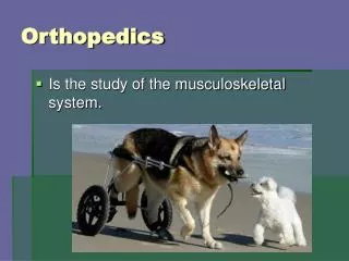
Mary ,a 4-year-old girl was brought in by her mother because of abnormal gait. The mother thinks that her daughter’s feet appeared to “turn in” when she walked.
History • Perinatal event • Development history including the motor development that may reveal cerebral palsy • The duration of the problem • Any family hx of gait problem
Parent’s concern • Pain, falling , limping • Sitting habits ‘W’ position • Any aggravating factors
Physical examination • Height and weight: any pathological causes • Spine: scoliosis • Neurological exam: musculoneurogical problem • Trendelenburg sign • Leg length • FROM of the hips, knees and ankles
The joint laxity • Flat foot • Gait and TORSIONAL PROFILE
What is your DDX ? What is the commonest cause of intoeing in a 4 yr old?
Physical examination Torsional profile • Foot progression angle • Forefoot alignment • Hip rotation • Thigh-foot angle
The foot progression angle of >5˚ intoeing at any age is abnormal • The tibia torsion measured by the thigh foot angle ranges from -30˚ to 30˚ • Tibia rotates laterally as growing • Measure the hip ext and int rotation for femoral torsion • Int hip rotation >70˚ means medial femoral torsion
Causes of in-toeing gait • Metatarsus adductus at birth/ infancy • Internal tibial torsion: toddler 12-24 mths • Increased femoral antevertion: 3-5 yrs
Metatarsus adductus • 1 in 1000 live births • Due to intrauterine packing • Adduction of forefoot with a convex lateral border • A function deformity and can be straightened
? Metatarsus Varus • This is to be differentiated from metatarsus adductus • This is a bony deformity with subluxation of the Tarsometatarsal jts • Cannot be brought into neutral position
? Club foot (talipes equinovarus) • Forefoot varus, • Heel varus and • Ankle equinus means point downwards can be fix or corrected by gentle pressure Exclude causes like CP. poliomyelitis
Metatarsus adductus Conservative Treatment • Stretching exercise during the first eight months of age • Performed 5 times at each diaper change Churgay CA. Dx and Tx of paediatric foor deformity. Am Fam Physician 1993;18:503-12
Metatarsus adductus Surgical treatment Surgical release of adductor hallucis tendon
Metatasus adductus Reasons for referral • Flexible deformity persists beyond 8 months of age • Rigid deformity
Metatasus adductus • 85-90% of metatarsus adducts identified at birth resolve without treatment by one year of age. Dietz FR. Intoeing. Am Fam Physician 1994;50:1249-59
Internal tibial torsion • Most common cause of in-toeing • Left side affected more than right • Male = female
Internal tibial torsion Cause • Intrauterine position • Sleeping in prone position • Sitting on feet
Internal tibial torsion Treatment • Avoid prone sleeping and sitting on feet • 90% gradually resolves on its own by 8 years of age. Salenius P. Rotational problem in children. Inst Course Lect 1994;13:429-39
Internal tibial torsion Reasons for referral • Persistent deformity beyond 1-2 year after the child learn walking • Significant or functional deformity • Thigh-foot angle of greater than 3 S.D. of mean • May need surgical correction if persisted beyond 8-10 yrs old
Increased femoral anteversion • Femur medially rotated on its long axis • Bilateral • Female > male
Increased femoral anteversion Causes • Familial • Sitting in a “W” position
Increased femoral anteversion Treatment • Nonoperative treatment is ineffective Salenius P. Rotational problem in children. Inst Course Lect 1994;13:429-39 Staheki LT. Torsion – treatment indication. Clin Ortho 1989; 247:61-6
Increased femoral anteversion Reasons for referral • Persistent deformity beyond 8 years of age • Significant cosmetic and functional disability • Anteversion in excess of 50 degree • Deformity more than 3 S.D. beyond the mean
Out-toeing • The cause is similar but opposite to those of in-toeing • Physiological when child learn to walk in infancy • Resolves by about 18 mths
Flat feet (Pes planus) • Loss of medial longitudinal arch • Physiological in infancy
Flat feet Causes • Joint laxity • Congenital vertical talus • Cerebral palsy, muscular dystrophy • Juvenile chronic arthritis
Flat feet Conservative treatment if • Pain free • Mobile and medial arch restored on dosiflexion of hallus or standing on tip toe
The shoes may be altered to raised the inner side of the heel • Placing an arch support inside the shoes
Flat feet Reasons for referral • Pain and stiff flat foot
A 2 year old boy was brought in by mother for URI symptoms. You notice bow legs of the child. The mother said he did not trip or fall. Boy’s ht was along 50 percentile but wt was 95th percentile for height.
What is the angular deformities? • Bow leg vs knock knee • Physiological vs pathological ? • How to assess the conditions?
Normal development • First 2 years of life : bow leg • Bowing increase till 2yrs old • Knock knee then develop between 3-4 yrs old • Knock knee resolves in 5-7 yrs to a normal adult alignment of 5 degree valgus
Pathological bow or knock knees • Asymmetry • Not consistent with the physiological sequences • Short stature • Severe deformity: >10cm of the intercondylar distance and > 10 cm of the intermalleolar distance • Rapid progression • Other musculoskeletal problem
History • Similar as in torsional problem • The chief complaint • AN, birth hx, development, health condition • Progression of symptoms • Family hx • Concern • Walking affected • Diet
Physical examination • Growth parameters • Spine, Hip, knee, Trendelenburg’s test • Neurological exam • Torsional profile • Intercondylar or intermalleolar distance
Intercondylar distance • Measure the degrees of bow leg with the medial malleoli touching. • Femoral tibial angle is a better assessment though difficult to obtain
Intermalleolar distance • Measures the knock knee • Distance between the medial malleoli and with the medial femoral condyles touching • Limit by the child’s size
Common causes of bow leg • Physiological • Rickets • Blounts disease • Achondroplasia • Trauma, infection,
Blount’s disease • Grow disturbance of the posterior medial aspect of the proximal tibia • Infantile form affects child of 1-3 years old • Bilateral • Obese • Early walkers • Usually in Africans
A sharp angulation of the proximal tibia • Spontaneous resolution is rare and need corrective surgery
Rickets • Inadequate mineralization • Vit D def • Nutritional • Vit D resistant • Renal osteodystrophy
Signs • SHORT stature • Poor muscles tone • Angular deformities of jts ( rosary beans) • Early onset causes bow legs • Late onset causes knock knees
DO Follow Up your patients for the physiological cases • And reassure the parents