Download
1 / 47
470 likes | 638 Views
Will Size Define a New Generation of Light Sources ?. David E. Moncton. Future Light Sources Conference JLab March 5, 2012. The most substantial societal impact of x-rays has come from small scale sources—x-ray tubes (14 Nobel prizes)

E N D
Will Size Define a New Generation of Light Sources? David E. Moncton Future Light Sources Conference JLab March 5, 2012
The most substantial societal impact of x-rays has come from small scale sources—x-ray tubes (14 Nobel prizes) Synchrotron radiation has made enormous impact in two ways Science accomplished (4 Nobel prizes) Development of extremely powerful methods based on high brilliance made possible by incoherent emission from relativistic beams X-ray free-electron lasers will also have enormous impact Access the fs timescale for the first time with significant flux Enable the development of methods exploiting the high peak brilliance made possible by coherent electron emission from relativistic beams Future machine beyond the ultimate single-mode FELs will combine the features of these three generations of sources Small scale, relativistic e-beams, and coherent emission. Summary
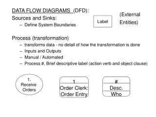
X-ray Science Driven by Beam Brilliance X-ray Lasers Coherent Emission Synchrotron Radiation Relativity X-ray Tubes
The Modern X-ray Tube Rigaku FR-E flux on crystal ~ 4 x 10⁹ ph/sec
Spiral CT (Computed Tomography) Imaging Note: We now know that much better images can be obtained by phase contrast methods, but are not possible with the cathode ray tube
X-ray Science Driven by Beam Brilliance X-ray Lasers Coherent Emission Synchrotron Radiation Relativity X-ray Tubes
Thousands of Protein Structures from One APS Beamline The Structural Biology Center APS Kappa Goniostat, ROBAC, Q315 detector 10 Tb NSF Storage
ATP-synthase structure (Boyer, Walker) 2003 Ion channels (Agre, MacKinnon) 2006 Eukaryotic transcription (Kornberg) 2009 Ribosime structure (Ramakrishnan, Steitz, Yonath) Nobel Prizes for Synchrotron X-ray Research
X-ray Science Driven by Beam Brilliance X-ray Lasers Coherent Emission Synchrotron Radiation Relativity X-ray Tubes
New Facilities Based on SASE Radiation SLAC Stanford
A Tutorial on Peak Brilliance • New scientific frontiers require access to the spatial and temporal regimes where new properties emerge that are out of reach with 3rd gen sources • This challenge requires the highest collimation, the smallest beam size, the shortest pulses and the best energy resolution—often simultaneously Peak Brilliance = # photons/Dx Dqx Dy Dqy Dt DE • The denominator is the phase space volume of the source which can be as small as the uncertainty principle allows—one photon mode • Therefore peak brilliance is a direct measure of the # photons in the most highly occupied mode—aka photon degeneracy • To convert from peak brilliance to photon degeneracy divide by 2.3 x 10²¹/ l3 (nm)
Peak Brilliance Example—The Advanced Photon Source with peak brilliance about 1023 at l = 0.1 nm 1023 = 0.04 Degeneracy = 2.3 x 10²¹/ l3 (nm) In other words the most highly occupied mode has, on average, a 4 percent chance of having one photon in it.
Peak Brilliance Example—The LCLS with peak brilliance about 1034 at l = 0.1 nm 2 x 1033 ~109 Degeneracy = 2.3 x 10²¹/ l3 (nm) What is the difference between sources with degeneracy of 109 and those sources with degeneracy from .01—100? It is the fundamentally different process producing the radiation– coherent vs incoherent emission, laser vs. light bulb Ring sources produce photons one electron at a time, proportional to the number N of electrons Lasing is a coherent electron process proportional to N2
Two Key Questions Are SASE sources the ultimate in x-ray technology? While SASE sources benefit from exponential gain, the resulting beam has poor quality compared to a single mode optical laser. No Are billion dollar central facilities the only option? Advances in accelerator, laser, and nano technology offer opportunities to build high performance x-ray sources of the scale of a university laboratory No
The SASE radiation is powerful, but noisy! Dw/w (%) t (fs) SASE Amplifies Random Electron Density Modulations Solution: Impose a strong coherent modulation with an external laser source
A Transform-Limited X-ray Pulse • Satisfies the Uncertainty Principle in all dimensions Transverse phase space Dx , Dy ~ 50 microns Dkx , Dky ~ 10-5 nm-1 Dx Dkx = ½ Dy Dky= ½ Longitudinal phase space Dt ~ 1fs - 1ps Dw ~ 2eV - 2meV Dw Dt = ½ A 1 milli-Joule pulse contains 5.0 x 10¹¹ 12 keV photons 3rd Gen: 1025 Peak brilliance = 1036
e- output Laser 266 nm 800 nm Modulator Buncher Radiator High Gain Harmonic Generation (HGHG) HGHG • Suppressed SASE noise • Amplified coherent signal • Narrowed bandwidth • Shifted wavelength SASE x105 Brookhaven Laser Seeding Demonstration L.H. Yu et al., Phys. Rev. Lett. 91, 74801 (2003).
X-ray Science Driven by Beam Brilliance X-ray Lasers Coherent Emission Synchrotron Radiation Relativity X-ray Tubes
There is a growing and urgent need to fill the large gap between laboratory x-ray tubes, and large central facilities. • At nano-centers such as EMSL that are not at synchrotron sources, advanced x-ray capability is the most important missing probe of matter. • Advances in accelerator and laser technology make high-brilliance, short pulses sources possible and affordable. Compact X-ray Sources ?
The Technical Opportunity Inverse Compton Scattering • Head-on collision between a relativistic electron and a photon e- lL energy = Ee= gme q lX • Normal Compton Scattering the photon has higher energy than the electron • The inverse process has the Thomson cross-section when Ee e X • The scattered photon satisfies the undulator equation with period lL/2 for head-on collisions (1+g2q2) lX = lL 4g2 • Therefore, the x-ray energy decreases by a factor of 2 at an angle of 1/g
Lyncean Technologies Compact Source Concept Courtesy of Ron Ruth Parameters of Source Average flux 109 photons/sec Source size 50 microns
photons/mrad2/eV photons/mrad2/eV 10 100 10 1 1 0.1 0.1 0.01 0.01 Full Calculations of the Photon Spectrum Normalized emittance = 1.0 mm Normalized emittance = 0.3 mm Note: Plane Perpendicular to Laser Polarization
Intensity Profile of 12 keV X-rays with 0.1% bw Calculations by Winthrop Brown, MIT LL ~1012photons/sec @ 100 MHz in 0.1% BW
1 kW cryo-cooled Yb:YAG drive laser X-ray beamline 2 Coherent enhancement cavity with Q=1000 giving 1 MW cavity power X-ray beamline 1 Inverse Compton scattering X-ray beamline 3 6 m Superconducting RF photoinjector 10 kW beam dump Electron beam of 1-100 mA average current at 10-30 MeV Superconducting RF Linac X-ray beamline 4 8 m Conceptual Multi-User Facility based on ICS
X-ray Science Driven by Beam Brilliance X-ray Lasers Coherent Emission Coherent ICS Synchrotron Radiation Incoherent ICS Relativity X-ray Tubes
How can coherent emission be achieved to boost ICS performance? Coherent ICS Answer: Maybe—by structuring the electron beam, not by laser seeding, but by using a structured nanocathode as a electron source, after T. Akinwande, MIT.
Coherent ICS It appears possible to manipulate these structured bunches in magnet arrays to compress, rotate, and exchange transverse and longitudinal coordinates, resulting in a sub nm periodic structure which will then coherently emit a hard x-ray beam. See talk by Bill Graves on Wednesday
20 cm 2 mm Asymmetric Ge (111) or Si (111) 10 µ Multilayer Osmic Condenser 30 µ 10 mrad Multilayer Osmic Collimator ‹ 100 µrad 3.3 mrad 60 cm │← 160 mm →│ Protein Crystallography Small Crystals with Fixed Wavelength and MAD • Goal: Achieve ICS images with 10 mm crystals of equal or better quality compared to rotating anode (Rigaku FR-E) with 100 mm crystals. Ge(111); DE = 16 eV; R = 67% Fixed Wavelength: Si (111); DE = 7 eV; R = 80% MAD:
Medical Imaging—Improved Absorption Images Current radiographs use 5-75 keV Bremstahlung spectrum Low energy range causes skin dose, no contrast High portion cause tissue dose with low contrast Only the range of energies around 30 keV useful ICS spectrum is ideal at 30 keV with 5-15% bandwidth Image quality improved and dose reduced We would establish collaboration with local radiologists to further study these factors in detail Medical Imaging/Therapy—Tuning to specific wavelengths Iodine contrast agent for blood imaging Gd or Pt based cancer therapy Medical Applications
Medical Imaging—Phase Contrast Method Few micron circular source is ideal Many milli-radian divergence illuminates large objects in short distance Few percent bandwidth can be fully utilized and presents no limitation Would improve medical imaging for soft tissue while reducing dose Could also utilize the single-shot mode for time-resolved images Simplest approach requires no optics But optics could reduce spot size, increase coherence, and increase illuminated area Medical Applications
Pico-second Science Synchrotron sources have 50-100 ps pulse lengths ICS source may have pulse lengths down to 100 fs At 1 ps the single-shot flux could be >109 photons in a 6% bandwidth Large bandwidths are appropriate for Laue method as pioneered by Wulff and co-workers at ESRF ICS could be run in a kHz mode for repetitive experiments Both diffraction and PC imaging modes possible Flux exceeds plasma sources by many orders of magnitude Flux exceeds storage ring pulse chopping schemes Would be a stepping stone to the big FELs Ultrafast Science Opportunities
Kirk Clark, Novartis Institutes for Biomedical Research Structural Biology is an integral component of many drug discovery programs. Guides medicinal chemistry efforts; turn-around time is critical Insight into protein function; novel structures benefit from tunable x-rays Epitope mapping for antibodies; rapid structures (even low resolution) valuable Benefits of bright, local x-ray source Time. Quick feedback on quality of small crystals and/or final datasets. Small crystals are more readily obtained with less reagents Reduced opportunity costs by avoiding needless improvements to good crystals. Facilitate expanding structural biology to integral membrane proteins. Costs. Reduced travel costs (currently traveling to Chicago/Zurich every 3 to 4 weeks), proprietary fees, access fees. Novartis 39
NIST Gaithersburg Campus Ultraviolet Synchrotron Radiation Laser Spectroscopy Advanced X-ray Facility SIRCUS Nanoscale Science & Technology CNST SURF III AXF NCNR Neutron Research
Louvre Museum VIS • LC2RMF = Laboratory of analytical chemistry and scientific imaging located in the Louvre • Objectives: Research, expertise and support for curators and conservators thanks to physico-chemical analyses. • Necessity of non destructive testing with high sensitivity because the studied materials are very complex • 1989: Installation of the accelerator AGLAE in the Louvre = ion beam analysis with an external microbeam (elemental analysis, direct on the artifacts) • 1997: Development of synchrotron radiation analysis = structural and molecular analysis but necessity of samples • 2009: Project of combination of AGLAE with an ICS in the Louvre for non destructive structural and elemental analysis (XRF, XANES, XRD) as well as for 3D imaging of works of art. X-rayUV
Virus Protein Science at the MIT STC • -M. Rossmann, Biological Sciences • Conduct structure determinations of proteins associated with viruses, and viral complexes with antibodies and antiviral agents. Extend current research efforts into • bacteriophages • animal RNA viruses • large ds DNA viruses • virus-membrane interactions • This Science & Technology Center could serve as a model for a future Purdue facility geared to on-site handling of viruses and pathogens difficult to handle at distant facilities. This would add to the new Hockmeyer Hall of Structural Biology and the Birck Nanotechnology Center at Purdue. • Picosecond Physics at the MIT STC • - S. Durbin, Physics Dept. • I. Laser pump deposits energy into the electronic system, and the lattice response can be tracked with an x-ray probe beam. • Use high rep rate (>106 Hz), ultrashort pulse length (0.1-5 ps, not available at ANY synchrotron source)) to observe • non-thermal melting, coherent phonons • ferroelectric/complex oxide transitions • porphyrin dynamics • II. X-ray pump, laser probe in semiconductors: Single 1 ps x-ray pulse (1010 x-rays) focused to 10 microns can generate up to 1017 cm-3 instantaneous deep core holes in a semiconductor. Laser monitor of absorption and reflectivity will reveal the quantum kinetic response of plasmons and phonons in this new many-body probe. Purdue University ds viruses Flaviviruses Bacteriophages Alpha viruses
University of Copenhagen Robert Feidenhans’l, Niels Bohr Institute, University of Copenhagen Absorption Phase shift Aim : Medical Imaging Franz Pfeiffer et al Lab source setup Cancerous Lymph node ESRF data
Within a decade FEL facilities will achieve single mode laser radiation in the hard x-ray regime with milli-Joule power levels. Within two decades superconducting linac-based facilities will have many such beamlines operating across a wide spectral range. There is no obvious “next generation” large facility. But…. Within a decade compact sources less than $10M will offer NSLS I level (2nd generation) x-ray properties. Within two decades compact sources will demonstrate coherent emission revolutionizing laboratory-scale capability. Large facilities always be the most powerful sources simply due to the physics of electromagnetic radiation from energetic charged particles. But the most creative science will most likely come from the laboratory sources, just as with laser sources today, and x-ray sources of the early 20th century. Conclusions (Predictions)