Download
1 / 3
30 likes | 101 Views
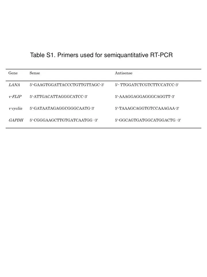
Table S1. Primers used for semiquantitative RT-PCR. 4. 4. 10. 10. 3. 3. 10. 10. 93.4. 96.6. 2. 2. 10. 10. 1. 1. 10. 10. 0. 0. 10. 10. 0. 1. 2. 3. 4. 0. 1. 2. 3. 4. 10. 10. 10. 10. 10. 10. 10. 10. 10. 10.

E N D
4 4 10 10 3 3 10 10 93.4 96.6 2 2 10 10 1 1 10 10 0 0 10 10 0 1 2 3 4 0 1 2 3 4 10 10 10 10 10 10 10 10 10 10 Fig. S1. Representative flow cytometric profiles with the ratio of mouse and human macrophages after the induction of macrophages as described in Material and methods. Human macrophages Mouse peritoneal macrophages CD56 CD49b CD11b CD14
Fig. S2. Anti-CD47 Ab did not inhibit the proliferation of PEL cells. PEL cell lines (BCBL-1, BC-2 and BCP-1) were incubated with 0, 0.3, 1, 3, 10 µg/ml anti-CD47 Ab for 72 h. A cell proliferation assay was carried out using MTT as described in Material and methods. Cell viability(%) Anti-CD47 Ab (μg/ml)