Download
1 / 50
500 likes | 663 Views
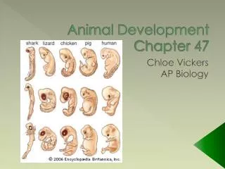
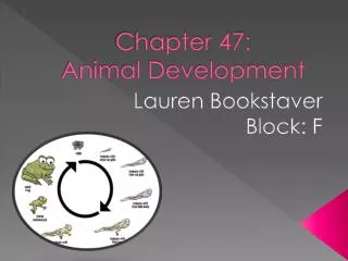
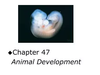
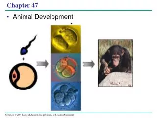
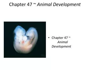
Chapter 47 Animal Development. Overview: A Body-Building Plan for Animals. From egg to organism, an animal’s form develops gradually. . The question of how a zygote becomes an animal has been asked for centuries.

E N D
From egg to organism, an animal’s form develops gradually • . • The question of how a zygote becomes an animal has been asked for centuries. • As recently as the 18th century, the prevailing idea was preformation, the notion that an egg or sperm contains an embryo that is a preformed miniature adult. • The competing theory is epigenesis, proposed 2,000 years earlier by Aristotle. • According to epigenesis, the form of an animal emerges from a relatively formless egg. • As microscopy improved in the 19th century, biologists could see that embryos took shape in a series of progressive steps. • Epigenesis displaced preformation as the favored explanation among embryologists.
Both preformation and epigenesis have some legitimacy. • Although the embryo’s form emerges gradually as it develops, aspects of the developmental plan are already in place in the eggs of many species. • An organism’s development is primarily determined by the genome of the zygote and also by differences that arise between early embryonic cells. • These differences set the stage for the expression of different genes in different cells. • In some species, early embryonic cells become different because of the uneven distribution within the unfertilized egg of maternal substances called cytoplasmic determinants. • These substances affect development of the cells that inherit them during the early mitotic divisions of the embryo.
In other species, the differences between cells are due to their location in the developing embryo. • Most species establish differences between early embryonic cells by a combination of these two mechanisms. • As development continues, selective gene expression leads to cell differentiation, the specialization of cells in structure and function. • Along with cell division and differentiation, development involves morphogenesis, the process by which an animal takes shape.
Concept 47.1 After fertilization, embryonic development proceeds through cleavage, gastrulation, and organogenesis
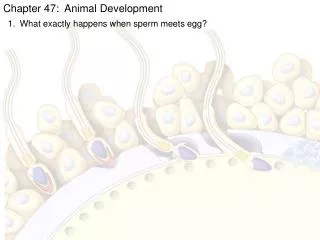
Fertilization activates the egg and brings together the nuclei of sperm and egg. • The gametes (egg and sperm) are both highly specialized cell types. • Fertilization combines haploid sets of chromosomes from two individuals into a single diploid cell, the zygote. • Another key function of fertilization is activation of the egg. • Contact of the sperm with the egg’s surface initiates metabolic reactions within the egg that trigger the onset of embryonic development. • Sea urchin fertilization has been extensively studied. • Sea urchin egg and sperm encounter each other after the animals release their gametes into seawater.
Fertilization activates the egg and brings together the nuclei of sperm and egg. • The jelly coat of the egg attracts the sperm, which swims toward the egg. • When the head of the sperm comes into contact with the jelly coat, the acrosomal reaction is triggered, and the acrosome, a specialized vesicle at the tip of the sperm, discharges its contents by exocytosis. • Hydrolytic enzymes enable the acrosomal process to penetrate the egg’s jelly coat. • The tip of the acrosomal process adheres to special receptor proteins on the egg’s surface. • These receptors extend through the vitelline layer, just external to the egg’s plasma membrane. • This lock-and-key recognition ensures that eggs will be fertilized only by sperm of the same species. • The sperm and egg plasma membranes fuse, and the sperm nucleus enters the egg’s cytoplasm. • Na+ channels in the egg’s plasma membrane open. • Na+ flows into the egg, and the membrane depolarizes, changing the membrane potential of the egg. • Such depolarization is common in animals.
Fertilization activates the egg and brings together the nuclei of sperm and egg. • Occurring within 1–3 seconds after the sperm binds to the egg, depolarization prevents additional sperm from fusing with the egg’s plasma membrane. • This fast block to polyspermy prevents polyspermy, the fertilization of the egg by multiple sperm. • Fusion of egg and sperm plasma membranes triggers a signal-transduction pathway. • Ca2+ from the egg’s endoplasmic reticulum is released into the cytosol and propagates as a wave across the fertilized egg. • High concentrations of Ca2+ cause cortical granules to fuse with the plasma membrane and release their contents into the perivitelline space, the space between the plasma membrane and the vitelline layer.
Fertilization activates the egg and brings together the nuclei of sperm and egg. • The vitelline layer separates from the plasma membrane. • An osmotic gradient draws water into the perivitelline space, swelling it and pushing it away from the plasma membrane. • The vitelline layer hardens into a fertilization envelope, which resists the entry of additional sperm. • The fertilization envelope and other changes in the egg’s surface function together as a long-term slow block to polyspermy. • The plasma membrane returns to normal, and the fast block to polyspermy no longer functions.
Fertilization activates the egg and brings together the nuclei of sperm and egg. • High concentrations of Ca2+ in the egg stimulate an increase in the rates of cellular respiration and protein synthesis, activating the egg. • Unfertilized eggs can be activated artificially by the injection of Ca2+ or by a variety of mildly injurious treatments, such as temperature shock. • It is even possible to activate an egg that has had its nucleus removed. • Evidently, proteins and mRNAs present in the cytoplasm of the unfertilized egg are sufficient for egg activation. • As the metabolism of the activated egg increases, the sperm nucleus swells and merges with the egg nucleus, creating the diploid nucleus of the zygote. • DNA synthesis begins and the first cell division occurs about 90 minutes after fertilization.
Fertilization in terrestrial animals, including mammals, is generally internal. • Secretions in the mammalian female reproductive tract alter certain molecules on the surface of sperm cells and increase sperm motility. • The mammalian egg is surrounded by follicle cells also released during ovulation. • A sperm must migrate through a layer of follicle cells before it reaches the zonapellucida, the extracellular matrix of the egg. • Binding of the sperm cell to a receptor on the zonapellucida induces an acrosomal reaction similar to that seen in the sea urchin. • Enzymes from the acrosome enable the sperm cell to penetrate the zonapellucida and bind to the egg’s plasma membrane.
The binding of the sperm cell to the egg triggers changes within the egg, leading to a cortical reaction, the release of enzymes from cortical granules to the outside via exocytosis. • The released enzymes catalyze alteration of the zonapellucida, which functions as a slow block to polyspermy. • The entire sperm, tail and all, enters the egg. • A centrosome forms around the centriole that acted as the basal body of the sperm’s flagellum. • This centrosome duplicates to form the two centrosomes of the zygote. • These will generate the mitotic spindle for the first cell division.
The envelopes of both egg and sperm nuclei disperse. • The chromosomes from the two gametes share a common spindle apparatus during the first mitotic division of the zygote. • Only after the first division, as diploid nuclei form in the two daughter cells, do the chromosomes from the two parents come together in a common nucleus. • Fertilization is much slower in mammals than in the sea urchin. • The first cell division occurs 12–36 hours after sperm binding in mammals
Cleavage partitions the zygote into many smaller cells. • A succession of rapid cell divisions called cleavage follows fertilization. • During this period, cells go through the S (DNA synthesis) and M (mitosis) phases of the cell cycle but may skip the G1 and G2 phases. • As a result, little or no protein synthesis occurs. • The first five to seven divisions form a cluster of cells known as the morula. • A fluid-filled cavity called the blastocoel forms within the morula, which becomes a hollow ball of cells called the blastula. • The zygote is partitioned into many smaller cells called blastomeres. • Each blastomere contains different regions of the undivided cytoplasm and, thus, may contain different cytoplasmic determinants. • Most animals have both eggs and zygotes with a definite polarity. • Thus, the planes of division follow a specific pattern relative to the poles of the zygote. • Polarity is defined by the heterogeneous distribution of substances such as mRNA, proteins, and yolk.
Yolk is most concentrated at the vegetal pole and least concentrated at the animal pole. • In amphibians, a rearrangement of the egg cytoplasm occurs at the time of fertilization. • The plasma membrane and cortex rotate toward the point of sperm entry. • The gray crescent is exposed, marking the dorsal surface of the embryo. • Molecules in the vegetal cortex are now able to interact with inner cytoplasmic molecules in the animal hemisphere, leading to the formation of cytoplasmic determinants that will later initiate development of dorsal structures. • Thus, cortical rotation establishes the dorsal-ventral (back-belly) axis of the zygote.
In frogs, the first two cleavages are vertical and result in four blastomeres of equal size. • The third division is horizontal, producing an eight-celled embryo with two tiers of four cells. • The unequal division of yolk displaces the mitotic apparatus and cytokinesis toward the animal end of the dividing cells in equatorial divisions. • As a result, animal blastomeres are smaller than those in the vegetal hemisphere. • Continued cleavage produces a morula and then a blastula. • Because of unequal cell division, the blastocoel is located in the animal hemisphere.
Animals with less yolk (such as the sea urchin) also have an animal-vegetal axis. • However, the blastomeres are similar in size, and the blastocoel is centrally located. • Yolk has its most pronounced effect on cleavage in the eggs of reptiles, many fishes, and insects. • The yolk of a chicken egg is actually an egg cell, swollen with yolk nutrients. • Cleavage of a fertilized bird’s egg is restricted to a small disk of yolk-free cytoplasm, while yolk remains uncleaved. • The incomplete division of a yolk-rich egg is meroblastic cleavage. • It contrasts with holoblastic cleavage, the complete cleavage of eggs with little or moderate yolk. • Early cleavage in a bird embryo produces a cap of cells called the blastoderm, which rests on undivided egg yolk.
The blastomeres sort into upper and lower layers, the epiblast and the hypoblast. • The cavity between these two layers is the avian version of the blastocoel. • This stage is the avian equivalent of the blastula. • In insects, the zygote’s nucleus is located within the mass of yolk. • Cleavage begins with the nucleus undergoing mitotic divisions, unaccompanied by cytokinesis. • These mitotic divisions produce several hundred nuclei, which migrate to the outer edge of the embryo. • After several more rounds of mitosis, plasma membranes form around each nucleus, and the embryo, the equivalent of a blastula, consists of a single layer of 6,000 cells surrounding a mass of yolk.
Gastrulation rearranges the blastula to form a three-layered embryo with a primitive gut. • Gastrulation rearranges the embryo into a triploblasticgastrula. • The embryonic germ layers are the ectoderm, the outer layer of the gastrula; the mesoderm, which fills the space between ectoderm and endoderm; and the endoderm, which lines the embryonic gut. • Sea urchin gastrulation begins at the vegetal pole where individual cells detach from the blastula wall and enter the blastocoel as migratory mesenchyme cells. • The remaining cells flatten to form a vegetal plate that buckles inward in a process called invagination. • The buckled vegetal plate undergoes extensive rearrangement of its cells, transforming the shallow invagination into a primitive gut, or archenteron. • The open end, the blastopore, will become the anus. • An opening at the other end of the archenteron will form the mouth of the digestive tube.
Frog gastrulation produces a triploblastic embryo with an archenteron. • Where the gray crescent was located, invagination forms the dorsal lip of the blastopore. • Cells on the dorsal surface roll over the edge of the dorsal lip and into the interior of the embryo, a process called involution. • Once inside the embryo, these cells move away from the blastopore and become organized into layers of endoderm and mesoderm, with endoderm on the inside. • As the process is completed, the lip of the blastopore encircles a yolk plug.
Gastrulation in the chick is similar to frog gastrulation in that it involves cells moving from the surface of the embryo to an interior location. • In birds, the inward movement of cells is affected by the large mass of yolk. • All the cells that will form the embryo come from the epiblast. • During gastrulation, some epiblast cells move toward the midline of the blastoderm then detach and move inward toward the yolk. • These cells produce a thickening called the primitive streak, which runs along what will become the bird’s anterior-posterior axis. • The primitive steak is the functional equivalent of the frog blastopore. • Some of the inward-moving epiblast cells displace hypoblast cells and form the endoderm. • Other epiblast cells move laterally into the blastocoel, forming the mesoderm. • The epiblast cells that remain on the surface form ectoderm. • The hypoblast is required for normal development and seems to help direct the formation of the primitive streak. • Some hypoblast cells later form portions of the yolk sac.
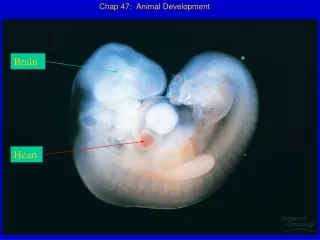
In organogenesis, the organs of the animal body form from the three embryonic germ layers. • Various regions of the three embryonic germ layers develop into the rudiments of organs during the process of organogenesis. • While gastrulation involves mass cell movements, organogenesis involves more localized morphogenetic changes in tissue and cell shape. • The first organs to form in the frog are the neural tube and notochord. • The notochord is formed from dorsal mesoderm that condenses above the archenteron. • Signals sent from the notochord to the overlying ectoderm cause that region of notochord to become neural plate. • This process is often seen in organogenesis: one germ layer signaling another to determine the fate of the second layer. • The neural plate curves inward, rolling itself into a neural tube that runs along the anterior-posterior axis of the embryo. • The neural tube becomes the brain and spinal cord.
Unique to vertebrate embryos is a band of cells called the neural crest, which develops along the border where the neural tube pinches off from the ectoderm. • Neural crest cells migrate throughout the embryo, forming many cell types. • Some have proposed calling neural crest cells the “fourth germ layer.” • Somites form in strips of mesoderm lateral to the notochord. • The somites are arranged serially on both sides along the length of the notochord. • Mesenchyme cells migrate from the somites to new locations. • The notochord is the core around which the vertebrae form. • Parts of the notochord persist into adulthood as the inner portions of vertebral disks. • Somite cells also form the muscles associated with the axial skeleton. • Lateral to the somites, the mesoderm splits into two layers that form the lining of the coelom.
As organogenesis progresses, morphogenesis and cell differentiation refine the organs that form from the three germ layers. • Embryonic development leads to an aquatic, herbivorous tadpole larva, which later metamorphoses into a terrestrial, carnivorous adult frog. • The derivatives of the ectoderm germ layer include epidermis of skin and its derivatives, epithelial lining of the mouth and rectum, cornea and lens of the eyes, the nervous system, adrenal medulla, tooth enamel, and the epithelium of the pineal and pituitary glands. • The endoderm germ layer contributes to the epithelial linings of the digestive tract (except the mouth and rectum), respiratory system, pancreas, thyroid, parathyroids, thymus, urethra, urinary bladder, and reproductive system. • Derivatives of the mesoderm germ layer are the notochord, the skeletal and muscular systems, the circulatory and lymphatic systems, the excretory system, the reproductive system (except germ cells), the dermis of skin, the lining of the body cavity, and the adrenal cortex.
Amniote embryos develop in a fluid-filled sac within a shell or uterus. • The amniote embryo is the solution to reproduction in a dry environment. • The shelled eggs of birds and other reptiles, as well as monotreme mammals, and the uterus of placental mammals provide an aqueous environment for development. • Within the shell or uterus, the embryos of these animals are surrounded by fluid within a sac formed by a membrane called the amnion. • Reptiles (including birds) and mammals are thus amniotes. • Amniote development includes the formation of four extraembryonic membranes: yolk sac, amnion, chorion, and allantois. • The cells of the yolk sac digest yolk, providing nutrients to the embryo. • The amnion encloses the embryo in a fluid-filled amniotic sac that protects the embryo from drying out. • The chorion cushions the embryo against mechanical shocks and works with the allantois to exchange gases between the embryo and the surrounding air. • The allantois functions as a disposal sac for uric acid and functions with the chorion as a respiratory organ.
Mammalian development has some unique features. • The eggs of most mammals are very small, storing little food. • Early cleavage is relatively slow in mammals. • In humans, the first division is complete after 36 hours, the second division after 60 hours, and the third division after 72 hours. • Relatively slow cleavage produces equal-sized blastomeres. • At the eight-cell stage, the blastomeres become tightly adhered to one another, causing the outer surface to appear smooth. • At completion of cleavage, the embryo has more than 100 cells arranged around a central cavity. • The blastocyst travels down the oviduct to reach the uterus. • Clustered at one end of the blastocyst is a group of cells called the inner cell mass that develops into the embryo and contributes to all the extraembryonic membranes.
The trophoblast, the outer epithelium of the blastocyst, secretes enzymes that break down the endometrium to facilitate implantation of the blastocyst. • The trophoblast thickens, projecting fingerlike projections into the surrounding maternal tissue, which is rich in vascular tissue. • Invasion by the trophoblast leads to erosion of the capillaries in the surrounding endometrium, causing the blood to spill out and bathe trophoblast tissue. • At the time of implantation, the inner cell mass forms a flat disk with an upper layer of cells, the epiblast, and a lower layer, the hypoblast. • As in birds, the human embryo develops almost entirely from the epiblast. • As implantation is completed, gastrulation begins. • Cells move inward from the epiblast through the primitive streak to form mesoderm and endoderm.
At the same time, extraembryonic membranes develop. • The trophoblast continues to expand into the endometrium. • The invading trophoblast, mesodermal cells derived from the epiblast, and adjacent endometrial tissue all contribute to the formation of the placenta. • The embryonic membranes of mammals are homologous with those of birds and other mammals. • The chorion, which completely surrounds the embryo and other embryonic membranes, functions in gas exchange. • The amnion encloses the embryo in a fluid-filled amniotic cavity. • The yolk sac encloses another fluid-filled cavity, which contains no yolk. • The yolk sac membrane of mammals is the site of early formation of blood cells, which later migrate to the embryo.
The fourth extraembryonic membrane, the allantois, is incorporated into the umbilical cord, where it forms blood vessels that transport oxygen and nutrients from the placenta to the embryo and rid the embryo of carbon dioxide and nitrogenous wastes. • The extraembryonic membranes of reptiles, where embryos are nourished with yolk, were conserved as mammals diverged in the course of evolution but with modifications adapted to development within the reproductive tract of the mother. • The completion of gastrulation is followed by the first events of organogenesis: the formation of the neural tube, notochord, and somites.
Concept 47.2 Morphogenesis in animals involves specific changes in cell shape, position, and adhesion
Morphogenesis is a major aspect of development in plants and animals, but only in animals does it involve cell movement. • Movement of parts of a cell can bring about changes in cell shape. • It can also enable a cell to migrate from one place to another within the embryo. • Changes in cell shape and cell position are involved in cleavage, gastrulation, and organogenesis. • Changes in the shape of a cell usually involve the reorganization of the cytoskeleton. • Consider how the cells of the neural plate form the neural tube. • First, the microtubules oriented parallel to the dorsal-ventral axis of the embryo help to lengthen the cells in that direction. • At the dorsal end of each cell is a parallel array of actin filaments oriented crosswise. • These contract, giving the cells a wedge shape that bends the ectoderm inward. • Similar changes in cell shape occur during other invaginations and evaginations of tissue layers throughout development.
The cytoskeleton is also drives cell migration. • Cells “crawl” within the embryo by extending cytoplasmic fibers to form cellular protrusions, in a manner akin to amoeboid movement. • The cellular protrusions of migrating embryonic cells are usually flat sheets (lamellipodia) or spikes (filopodia). • During gastrulation, invagination is initiated by the wedging of cells on the surface of the blastula, but the movement of cells deeper into the embryo involves the extension of filopodia by cells at the leading edge of the migrating tissue. • The cells that first move through the blastopore and along the inside of the blastocoel drag others along behind them as a sheet of cells. • This involuted sheet of cells forms the endoderm and mesoderm of the embryo.
Cell crawling is also involved in convergent extension, a type of morphogenetic movement in which the cells of a tissue layer rearrange themselves so the sheet converges and extends, becoming narrower but longer. • Convergent extension allows the archenteron to elongate in the sea urchin and frog and is responsible for the change in shape of a frog embryo from spherical to submarine shaped. • The movements of convergent extension probably involve the extracellular matrix (ECM), a mixture of secreted glycoproteins lying outside the plasma membrane. • ECM fibers may direct cell movement by functioning as tracks, directing migrating cells along particular routes. • Some ECM substances, such as fibronectins, help cells migrate by providing anchorage for crawling. • Other ECM substances may inhibit migration in certain directions.
In frog gastrulation, fibronectin fibers line the roof of the blastocoel. • As the future mesoderm moves into the interior of the embryo, cells at the free edge of the mesodermal sheet migrate along these fibers. • Researchers can prevent the attachment of cells to fibronectin (and prevent inward movement of the mesoderm) by injecting embryos with antifibronectin antibodies. • As migrating cells move along specific paths through the embryo, receptor proteins on their surfaces pick up directional cues from the immediate environment. • Such signals from the ECM can direct the orientation of cytoskeletal elements to propel the cell in the proper direction. • Cell adhesion molecules (CAMs), located on cell surfaces, bind to CAMs on other cells. • CAMs vary in amount and chemical identity with cell type. • These differences help to regulate morphogenetic movement and tissue binding. • Cadherins are also involved in cell-to-cell adhesion. • Cadherins require the presence of calcium for proper function. • There are many cadherins, and the gene for each cadherin is expressed in specific locations at specific times during embryonic development.
Concept 47.3 The developmental fate of cells depends on their history and on inductive signals
Development requires the timely differentiation of cells in specific locations. • Two general principles integrate the current understanding of the genetic and cellular mechanisms that underlie differentiation during embryonic development.
First, during early cleavage divisions, embryonic cells must somehow become different from one another. • In many animal species, initial differences result from uneven distribution of cytoplasmic determinants (mRNAs, proteins, and other molecules) in the unfertilized egg. • The resulting differences in the cytoplasmic composition of cells help specify body axes and influence the expression of genes that affect the developmental fate of cells. • For example, the cells of the inner cell mass are located internally in the early human embryo, while trophoblast cells are located on the outer surface of the blastocyst. • The difference in cell environment determines the fate of these cells.
Second, once initial cell asymmetries are set up, subsequent interactions among the embryonic cells influence their fate, usually by causing changes in gene expression. • This mechanism is termed induction. • Induction, which brings about the differentiation of many specialized cell types, is mediated by diffusible chemical signals or by cell-surface interactions.
Fate mapping can reveal cell genealogies in chordate embryos. • Fate maps illustrate the developmental history of cells. • In classic experiments in the 1920s, German embryologist Vogt charted fate maps for different regions of early amphibian embryos. • His work provided evidence that the lineage of cells making up the three germ layers created by gastrulation is traceable to cells in the blastula, before gastrulation begins. • Developmental biologists have combined fate-mapping studies with experimental manipulation of parts of embryos. • Two important conclusions have emerged. • “Founder cells” give rise to specific tissues in older embryos. • As development proceeds, a cell’s developmental potential (the range of structures it can form) becomes restricted.
The eggs of most vertebrates have cytoplasmic determinants that help establish the body axes. • A bilaterally symmetrical animal has an anterior-posterior axis, a dorsal-ventral axis, and left and right sides. • Establishing this basic body plan is a first step in morphogenesis and a prerequisite for the development of tissues and organs. • In frogs, locations of melanin and yolk define the animal and vegetal hemispheres respectively. • The animal-vegetal axis indirectly determines the anterior-posterior body axis. • Fertilization in frogs triggers cortical rotation, which establishes the dorsal-ventral axis and leads to the appearance of the gray crescent, whose position marks the dorsal side. • Once any two axes are established, the third (right-left) is specified by default. • Molecular mechanisms then carry out the program associated with that axis.
In amniotes, body axes are not fully established until later. • In chicks, gravity is involved in establishing the anterior-posterior axis as the egg travels down the oviduct before being laid. • Later, pH differences between the two sides of the blastoderm establish the dorsal-ventral axis. • In mammals, no polarity is obvious until after cleavage, although recent research suggests that the orientation of the egg and sperm nuclei before fusion may play a role in determining the axes. • In many species with cytoplasmic determinants, only the zygote is totipotent, capable of developing into all cell types found in the adult. • The fate of embryonic cells is affected by both the distribution of cytoplasmic determinants and cleavage pattern. • In frogs, the first cleavage occurs along an axis that produces two identical blastomeres with identical developmental potential.
The cells of the mammalian embryo remain totipotent until the 16-cell stage, when they become arranged into the precursors of the trophoblast and inner cell mass of the blastocyst. • At that time, location determines cell fate. • At the 8-cell stage, each of the blastomeres of the mammalian embryo can form a complete embryo if isolated. • The progressive restriction of potency is a general feature of development in animals. • In some species, the cells of the early gastrula retain the capacity to give rise to more than one kind of cell, although they are no longer totipotent. • In general, the tissue-specific fates of cells in the late gastrula are fixed. • Even if manipulated experimentally, they will give rise to the same type of cells as in a normal embryo.
Inductive signals play an important role in cell fate determination and pattern formation. • Once embryonic cell division creates cells that are different from one another, the cells begin to influence each other’s fates by induction. • At the molecular level, the effect of induction is usually the switching on of a set of genes that make the receiving cells differentiate into a specific tissue. • In the 1920s, Hans Spemann and Hilde Mangold carried out a set of transplantation experiments. • These experiments showed that the dorsal lip of the blastopore in an early gastrula serves as an organizer of the embryo by initiating a chain of inductions that results in the formation of the notochord, neural tube, and other organs. • Developmental biologists are working to identify the molecular basis of induction by Spemann’s organizer (also called the gastrula organizer or simply the organizer).
A growth factor called bone morphogenetic protein 4 (BMP-4) is active exclusively in cells on the ventral side of the amphibian gastrula. • BMP-4 induces those cells to form ventral structures. • Organizer cells inactivate BMP-4 on the dorsal side of the embryo by producing proteins that bind to BMP-4, rendering it unable to signal. • This allows formation of dorsal structures such as the notochord and neural tube. • Proteins related to BMP-4 and its inhibitors are also found in other animals, suggesting that they evolved long ago and may participate in development in many different organisms.
Many inductions involve a sequence of inductive steps that progressively determine the fate of cells. • In late gastrula of the frog, ectoderm cells destined to form the lenses of the eyes receive inductive signals from the ectodermal cells that will form the neural plate. • Later, inductive signals from the optic cup, an outgrowth of the developing brain, complete the determination of lens-forming cells. • Inductive signals play a major role in pattern formation, the development of an animal’s spatial information. • Positional information, supplied by molecular cues, tells a cell where it is relative to the animal’s body axes.
Limb development in chicks serves as a model of pattern formation. • Wings and legs of chicks begin as bumps of tissue called limb buds. • Each component of a chick limb develops with a precise location and orientation relative to three axes, the proximal-distal axis (shoulder-to-fingertip), the anterior-posterior axis (thumb-to-little-finger), and the dorsal-ventral axis (knuckle-to-palm). • A limb bud consists of a core of mesodermal tissue covered by a layer of ectoderm. • Two critical organizer regions are present in all vertebrate limb buds. • The cells of these regions secrete proteins that provide key positional information to the other cells of the bud.
One limb-bud organizer region is the apical ectodermal ridge (AER), a thickened area of ectoderm at the tip of the bud. • The AER is required for the outgrowth of the limb along the proximal-distal axis and for patterning along this axis. • The cells of the AER produce several secreted protein signals, belonging to the fibroblast growth factor (FGF) family. • These signals promote limb-bud outgrowth. • If the AER is surgically removed and beads soaked in FGF are put in its place, a nearly normal limb will develop. • The AER (and other limb-bud ectoderms) also appears to guide pattern formation along the limb’s dorsal-ventral axis. • If the ectoderm of the limb bud, including the AER, is detached from the mesoderm and rotated 180°back-to-front, the limb elements that form have reversed dorsal-ventral orientation.
The second major limb-bud organizer region is the zone of polarizing activity (ZPA), a block of mesodermal tissue located underneath the ectoderm where the posterior side of the bud is attached to the body. • The ZPA is necessary for proper pattern formation along the anterior-posterior axis of the limb. • Cells nearest the ZPA give rise to posterior structures (such as our little finger); cells farthest from the ZPA form anterior structures (such as our thumb). • Tissue transplantation experiments support the hypothesis that the ZPA produces an inductive signal that conveys positional information indicating “posterior.” • The cells of the ZPA secrete a protein growth factor called Sonic hedgehog. • If cells genetically engineered to produce large amounts of Sonic hedgehog are implanted in the anterior region of a normal limb bud, a mirror-image limb bud results. • Extra toes and fingers in mice (and maybe humans) result from the production of Sonic hedgehog in the wrong part of the limb bud.
We can conclude from these experiments that pattern formation requires cells to receive and interpret environmental cues that vary with location. • These cues tell cells where they are in the 3-D realm of a developing organ. • Organizers such as the AER and the ZPA function as signaling centers. • The AER and ZPA also interact with each other via signaling molecules and signaling pathways, to influence each other’s developmental fates. • What determines whether a limb bud develops into a forelimb or a hindlimb? • The cells receiving signals from the AER and ZPA respond according to their own developmental histories. • Earlier developmental signals have set up patterns of gene expression that distinguish future forelimbs from future hindlimbs. • Construction of a fully formed animal involves a sequence of events that include many steps of signaling and differentiation. • Initial cell asymmetries allow different types of cells to influence each other to express specific sets of genes. • The products of these genes direct cells to differentiate into specific types. • Coordinated with morphogenesis, various pathways of pattern formation occur in all the different parts of the developing embryo. • These processes produce a complex arrangement of multiple tissues and organs, each functioning in the appropriate location to form a coordinated organism.