Download
1 / 57
630 likes | 1.32k Views
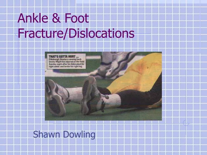
Ankle & Foot Fracture/Dislocations. Shawn Dowling. ANKLE 3 Primary Joints Medial malleolus w/medial talus Tibial plafond w/talar dome Lat malleolus w/lat talus 3 Bones: Tibia, Fibula and Talus. 3 sets of Ligaments: Lateral collaterals (ATFL, CFL, PTFL) Syndesmotic Ligaments

E N D
Ankle & Foot Fracture/Dislocations Shawn Dowling
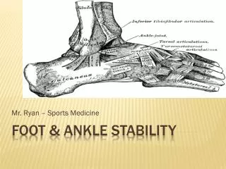
ANKLE 3 Primary Joints Medial malleolus w/medial talus Tibial plafond w/talar dome Lat malleolus w/lat talus 3 Bones: Tibia, Fibula and Talus 3 sets of Ligaments: Lateral collaterals (ATFL, CFL, PTFL) Syndesmotic Ligaments Medial collaterals (Deltoid) ANATOMY 101
BONES Fibula Tibia Talus
LIGAMENTS Syndesmotic Ligaments Medial Collateral Ligaments Lateral Collateral Ligaments
JOINTS Fibulotalar Tibiotalar (mid) Tibia Fibula Tibiotalar (lateral) Talus
Complicated (28 bones, 57 articulations) Subdivided in 3 segments & mvts Hindfoot - inv/ever Midfoot - abd/add Forefoot – flex/ext Joints Talo-crural jnt Inversion/eversion Hindfoot – mid foot (Choparts) Inversion/eversion Midfoot – forefoot (Lisfranc’s)* Abd/adduction MTP-IP Flex/extension FOOT
HINDFOOT talus calcaneus navicular cuboid MIDFOOT Medial cuneiforms metatarsals sesamoids FOREFOOT phalanges
Choparts Lisfrancs MTP IP
C E B A F D
What are stable fractures? • Ankle forms a ring • Disruption of only 1 structure is stable • Disruption of > 1 is unstable
Approach to Ankle/Foot X-rays • Go through complete approach (ABC’s) • 3 views- AP, lat, Mortise (15-20° int rot) ankle, • Direct evidence of injury: assess bones • Indirect evidence of injuries: are all ankle measurements normal? Joint effusion? • Describe x-ray, rather than simply naming it
Management • In general • Chip/avulsion #’s <3mm = Tx as sprain • Non-displaced, non-intra-articular, stable #’s • 3 wks NWB cast, 3-5 wks WB cast, f/u with cast clinic • Unstable #’s, intra-articular # - speak with Ortho • Open – saline soaked dsg, IV ABx, Td, Ortho urgently • NV compromise – reduce and call Ortho Urgently
Diagnosis?Classification?Treatment? Does it change you mgmt if they have a tender deltoid ligament?
Lateral Malleoli #’s • MC ankle #, MOI: usu inversion injury • Weber classification – used to determine risk of syndesmosis injury and therefore need for operative repair • Management • NWB x 3wks, WB x 3-5wks* • Refer B’s or C’s, Functional bimalleolar’s to ortho
Stable ? • Is the location significant? • Management? • What measurements/lines do you look at in the ankle? • What do they signify?
Syndesmosis injury 1 >10 mm B A Medial clear Space <5mm 2 3 4 A-B = talar tilt <3 is normal
Bimalleolar/Trimalleolar #’s • Involve the medial, lateral and/or posterior malleoli • Splint, pain control, NPO • Need to speak to ortho as they will likely need OR
PILON #Mechanism of injury- axial load? Associated injuries- calcaneus, C,T & L spine, pelvis, intra-abdominal.Management- OR, approx 50% are open fractures
Description? What do you want to know/assess?What do you want to do? How?
Ankle Dislocations • Relatively common, usually assoc w/# • Describe the position of foot/talus to tibia • If open, Tx as such • X-rays should not delay reduction if NV compromise or skin tenting present • Analgesia/PS, Reduce, splint, post-red films
Pediatric Ankle Injuries • Not just little adult # • The ligament attachments are stronger than the physis therefore more #’s, less sprains • Overall management is similar to adults • Although with fractures you can accept more angulation (little to no displacement) • LLC casts are the initial choice for most #’s
Can we apply OAR/OFR in children? • Six studies looked at validating OAR in peds • Different age groups (2-18, 6-16) • Sens 85*-100*% • Considered all # • Some considered all #, others only “significant #”)
BMJ 2003. Accuracy of OAR to exclude fractures of the ankle and mid-foot: A systematic review • This study references all of the OAR done in children as well as adults
Problems with the studies • Haven’t come up with a common definition of significant # • Unsure of what to do with SH-1, inconsistent Dx • Local practice (and Edmonton) – variable some apply it, some use rule + discretion, others use clinical judgement
Conclusion • This needs to be further studied • Need to determine which #’s are significant • But I think they will likely be validated • Although I think they’ll have to Tx SH1 as distinct injuries
Describe fracture? Classification? Management? SH-2 LLC x 3 wks, then SLC X 1-3 wks
Describe fracture? Classification? Management? SH2 Reduction/immbolize (air cast)
SALTR Lower (epiphysis) Ram (Crush) Straight Thru the Physis Above (metaphysis)
Non-operative management for SH 1-2 • Attempt closed reduction, can accept more angulation • Long leg cast x 3wks, followed by SLC x 3wks • SH 3-4 -> OR • SH 5 ->poor fx prognosis • Complications for SH 3-5 include growth arrest, limb length discrepancy
Acute Skin necrosis NV injury Compartment syndrome # Blisters Wound infection/osteo Chronic Mal-union Non-union Post-traumatic arthritis AVN Chronic pain Chronic instability Ankle # Complications
Describe # Do you need to speak to Ortho? ?ottawa ankle rules Talar Dome # Yes – Ortho to see in cast clinic
Describe # • Anything special about this bone? • Blood flow distal to proximal like scaphoid and proximal femur, therefore inc AVN risk • Is there a classification system for these #’s?
Talus Neck Body Head Chopart’s joint
Talar fractures • Minor talar fractures • Chip and avulsion fractures of neck ,head, and body. • Usually same mechanism as ankle sprains • Talar neck fractures • 50% of major talar injuries. • extreme dorsiflexion force • Hawkins classification • Talar body fractures • 23% of all talar fractures (including minor fractures) • Major talar body fractures are uncommon • usually axial loading (e.g. falls) • Talar head fractures • Uncommon (5-10%) • compressive force transmitted up through the talonavicular joint applied on a plantarflexed foot
Hawkins Classification of Talar Neck Fractures • Type 1: = nondisplaced; • Type 2: subtalar subluxation • Type 3: dislocation of the talar body (50% open #’s) • Type 4: dislocation of the talar body & distraction of the talonavicular joint. • Fracture type influences management & prognosis Thanks Moby
Describe injury. • Name this injury.v • Management?
Describe injury. • Name this injury • Lisfranc • Management? • OR
What to look for on x-ray: • Normally, medial aspect of metatarsals 1-3 should align with medial borders of cuneiforms • Metatarsals should be aligned dorsally with tarsals on lateral view • Medial 4th metatarsal should align with medial cuboid • Any fracture or dislocation of the navicular or cuneiforms or widening between metatarsals 1-3 • Proximal 2nd metatarsal # is pathognomonic Thanks Dave
Normal Lisfranc joint alignment • Tx: • Need to speak to ortho • May try closed reduction
Describe. • Management • NWB cast • # usu from direct trauma
Describe. • Management • Walking cast x 2-3 weeks • Avulsion type #
Metatarsal # Treatment: • Nondisplaced or min displaced fractures of metatarsal 2-4 stiff shoe, casting, or fracture brace. • Non displaced 1st metatarsal NWB BK walking cast (cuz it’s a major WB surface) • Displaced 1st or 5th metatarsal ER ortho • Attempt closed reduction if >3mm displacement or 10 degrees angulation Thanks Dave