Download
1 / 1
10 likes | 172 Views
A Surgical Light Device to Gauge Depth & Position of Surgical Cannula Catherine Augello, Hector Muñoz, Barbara Thorne-Thomsen & Michael Zhao Department of Bioengineering Rice University (TeamLANAR@gmail.com). RESULTS.

E N D
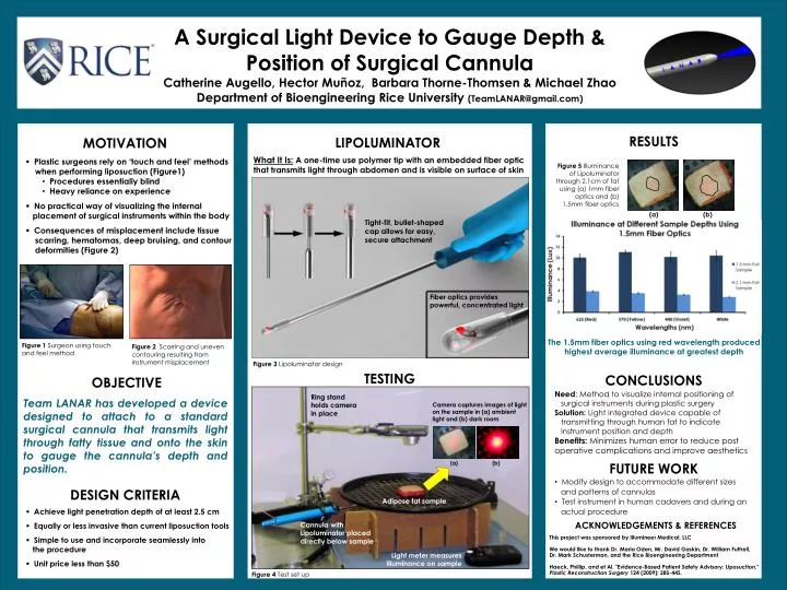
A Surgical Light Device to Gauge Depth & Position of Surgical Cannula Catherine Augello, Hector Muñoz, Barbara Thorne-Thomsen & Michael Zhao Department of Bioengineering Rice University (TeamLANAR@gmail.com) RESULTS LIPOLUMINATOR OBJECTIVE CONCLUSIONS What It Is: A one-time use polymer tip with an embedded fiber optic that transmits light through abdomen and is visible on surface of skin Figure 5 Illuminance of Lipoluminator through 2.1cm of fat using (a) 1mm fiber optics and (b) 1.5mm fiber optics Team LANAR has developed a device designed to attach to a standard surgical cannula that transmits light through fatty tissue and onto the skin to gauge the cannula’s depth and position. Need: Method to visualize internal positioning of surgical instruments during plastic surgery Solution: Light integrated device capable of transmitting through human fat to indicate instrument position and depth Benefits: Minimizes human error to reduce post operative complications and improve aesthetics FUTURE WORK (a) (b) • Modify design to accommodate different sizes • and patterns of cannulas • Test instrument in human cadavers and during an • actual procedure • Tight-fit, bullet-shaped cap allows for easy, secure attachment MOTIVATION • Plastic surgeons rely on ‘touch and feel’ methods • when performing liposuction (Figure1) • Procedures essentially blind • Heavy reliance on experience • No practical way of visualizing the internal • placement of surgical instruments within the body • Consequences of misplacement include tissue • scarring, hematomas, deep bruising, and contour • deformities (Figure 2) Fiber optics provides powerful, concentrated light The 1.5mm fiber optics using red wavelength produced highest average illuminance at greatest depth Figure 3 Lipoluminator design TESTING Ring stand holds camera in place Figure 1 Surgeon using touch and feel method Figure 2 Scarring and uneven contouring resulting from instrument misplacement DESIGN CRITERIA Camera captures images of light on the sample in (a) ambient light and (b) dark room • (b) • Achieve light penetration depth of at least 2.5 cm • Equally or less invasive than current liposuction tools • Simple to use and incorporate seamlessly into • the procedure • Unit price less than $50 ACKNOWLEDGEMENTS & REFERENCES Adipose fat sample This project was sponsored by Illumineer Medical, LLC We would like to thank Dr. Maria Oden, Mr. David Gaskin, Dr. William Futtrell, Dr. Mark Schusterman, and the Rice Bioengineering Department Haeck, Phillip, and et Al. "Evidence-Based Patient Safety Advisory: Liposuction." Plastic Reconstruction Surgery 124 (2009): 28S-44S. Cannula with Lipoluminator placed directly below sample Light meter measures illuminance on sample Figure 4 Test set up