Download
1 / 97
1k likes | 1.04k Views
Venous Disorders. Venous return. Muscle pump ( peripheral hearts) - ve intra thoracic pressure Arterial pulsation Vise at ergo. Varicose Veins. Dilated, elongated & tortuous vein of the LL problem comes from incompetent calve. 10 -20 % of worlds population have varicose veins.

E N D
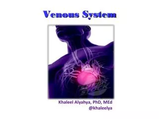
Venous return • Muscle pump ( peripheral hearts) • - ve intra thoracic pressure • Arterial pulsation • Vise at ergo
Varicose Veins • Dilated, elongated & tortuous vein of the LL • problem comes from incompetent calve. • 10 -20 % of worlds population have varicose veins.
Causes of varicos veins in lower limbs. • Secondary • Obstruction of venous outflow. • Pregnancy. • Fibroids • Ovarian cysts. • Abdominal lymphadenopathy • Pelvic cancer (cervical, uterus, ovary, rectum) • Ascites • Illiac vein thrombosis. • Retroperitoneal fibrosis • Valve destruction. • DVT • High flow and pressure: • Arteriovenous fistula ( esp the aquired traumatic variety)eg.Klippel-Trenaunay syndrome (which is one form of congenital AV malformation syndrome) • Primary: • Cause not known. Often familial.Probably weakness of vein wall that permits valve ring to dilate. • Congenital abscence of valves very rare.
Primary Hereditary Occupational Pregnancy obesity Secondary Venous obstruction Venous compression A/V fistula Varicose Veins (Etiology)
Varicose Veins (Etiology) • Obstruction of venous outflow. • Pregnancy. • Fibroids • Ovarian cysts. • Abdominal lymphadenopathy • Pelvic cancer (cervical, uterus, ovary, rectum) • Ascites • Illiac vein thrombosis. • Retroperitoneal fibrosis • Valve destruction. • DVT • High flow and pressure: • Arteriovenous fistula ( esp the aquired traumatic variety) • Klippel-Trenaunay syndrome (which is one form of congenital AV malformation syndrome
Primary V V History: young and middle aged women most commonly affected.1:10 men :women Aggrevating factors associated with increased incidence of varicose veins: female sex, parity, clothing, prolonged standing, marked obesity, Secondary V V History: Any age most commonly affect men Aggrevating factors associated with increased incidence of varicose veins: DVT Trauma Compression fracture Varicose Veins
Disfiguring effects of the veins usually principle complaint Pain, dull ache, and heaviness felt in calves and lower leg worse during day esp. on standing up, relieved by lying down for 15-30 min. Edema (swelling around ankle) Aggregated by standing Relived by recumbency night cramps Disfiguring effects of the veins usually principle complaint Pain, dull ache, and heaviness felt in calves and lower leg worse during day esp. on standing up and walking relieved by lying down for 15-30 min. Edema (swelling around ankle) Aggregated by standing Relived by recumbency night cramps Post phlebitic syndrome dilated veins, venous stars, pigmentation, eczema and ulceration. Varicose Veins (symptoms)
Dilated elongated tortuous veins Types of varices Tubular with dilated LSV or SSV Saccular incompetent perforator ( blow out) Signs of PPS - ve Special testes Modified perthe’s Cough impulse Trendelenburg’s test Multiple tourniquet test Shwartz test Dilated elongated tortuous veins Types of varices Serpintine dilated tributeries Spider indicate A/ V fistula Signs of PPS + ve Special testes Modified perthe’s Cough impulse Trendelenburg’s test Multiple tourniquet test Shwartz test Varicose Veins (signs)
LSV behind the knee LSV Vein of Leonardo ( post arch vein ) LSV behind the knee Vein of Leonardo ( post arch vein ) LSV in front of medial malleolus
Communicator just below knee LSV behind the knee LSV Vein of Leonardo ( post arch vein ) Vein of Leonardo ( post arch vein )
LSV starting at mid thigh Communicator and pass behind the knee mid thigh Communicator Antromedial and calf Group of tributaries Antromedial and calf Group of tributaries
Examination: • Inspection: • ask patient to stand up. • look for abnormal visible subcutaneous veins. if dilated and tortuous=varicose veins. • record size and shape of the veins. • venous stars (minute veins radiating from a single feeding vein • oedema • inspect skin esp. lower medial 1/3 for pigmentation, eczema, ulceration.
Examination: • Palpation: • feel along the course of the veins and feel the tension in the veins • feel saphenofemoral and saphenopopliteal junctions and ask patient to cough, a strong cough impulse indicates incompetent valves. • feel along medial side of lower leg for tender defects in deep facia with patient standing and lying, these are sites of incompetent valves. • look for pitting oedema, thickening, and tenderness. • brown pigmentation , eczema and ulceration.
Examination • Tourniquet tests. • to check for the site of the incompetent valves. • Lie patient flat and elevate one leg • place tourniquet along upper 1/3 of thigh • ask patient to stand up • if veins fill above tourniquet the incompetence above. • Trendelenburgs test. • direct digital pressure on long saphenous vein valve. • patient first lying with leg up • stand up patient
Examination • Percussion: • transmission of percussion waves downward implies incompetent valves ( Shwartz test). • Place fingers of one hand on lower limit of visible vein and tap top. • Auscultation: • listen over clusters of veins especially if they remain distended when patient lies down may be arteriovenous fistula.
Examination • General examination: • examine abdomen, incl rectal and vaginal examination. • men: palpate testes, testicular tumours can be small but cause massive enlargement of the abdominal nodes with vena caval obstruction. • look for dilated collateral veins on abdomen. • direction of flow: Harvey's test (emty veins with 2 fingers and see where it fills from)
Investigation • Routine Lab mainly BSL • Hand held Doppler Continuous wave Doppler (CWD) (phono-angiography)
Investigation • Doppler US
Investigation Duplex US gold standard • (B mode ultrasound and a coupled doppler probe) • allows direct visualiastion of veins, direction of flow can be recorded
Investigation • Plethysmography and Venography are obsolete • Venous pressure • Radio-active isotope scanning • Arteriograpgy if A/V fistula
Complications: • Haemorrhage • Oedema • Skin pigmentation • Lipodermatosclerosis • Varicose eczema • Venous ulceration • Thrombophlebitis • Atrophie blanche • Marjolin ulcer • Equinous deformity
Varicose eczema Thrombophelbitis
Treatment • A. Non- operative management. • walking should be encouraged and prolonged sitting and standing should be forbidden • patient should elevate leg as frequently as possible to reduce venous pressure. • elastic stockings. extending from distal metatarsals to just below the knee
Treatment Compression sclerotherapy. • permanent fibrotic occlusion of collapsed veins. • patient is recumbent and veins collapsed, • a small amout of 0.5 ml of sclerosing solution (3% sodium tetradecyl sulfate) is injected into each varix
Compression sclerotherapy. • continuous pressure is maintained for 1-2 weeks with elastic stockings. • much less expensive than surgery • if successful it gives the best cosmetic result. • long term results are worse than surgery. • best for small unsightly veins, dilated superficial veins, lower leg perforators, and recurrent or persistant veins after surgery • unsatisfactory at or above the knee
Endo-venous laser • Peri-venous LA • 810 nm diode • Time consuming • Less painful
Radiofrequency ablation • Peri-venous LA/ regional anaesthesia • Pode expansion in CFV • Cook at 85oC • Time consuming
Treatment Surgical therapy. • Indications: • severe symptoms • very large varices • attacks of superficial phlebitis • haemorrhage from rupturd varices. • ulceration from venous stasis. • cosmetic reasons.
Treatment Surgical therapy. • identify all perforating and superficial veins preoperatively and mark them. • results depend on thoroughness of the procedure. • postoperatively leg is supported with elastic bandages for approximately 6 weeks. • elevation of leg in bed minimizes postop swelling. • recurrens rate of about 10%. most common cause is failure to ligate all the tributaries, and incompetent perforators.
External valvular stent Adjustable gore-tex/ dacron cuff ?physiological
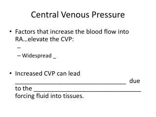
Deep Vein Thrombosis • Only 1/3 of DVT's cause symptoms and signs. • predisposition to thrombosis is predicted with Virchow's triad. • Change in vessel wall; distention, injury, inflammation, trauma. • Diminished rate of blood flow; during and after operations (postop rare before 40years, most common operations;obesity, operations for cancer,prostate and hip), debilitating diseases • Increased coagulability of the blood; infections, after haemorrhage, visceral cancers,during pregnancy, hypercoagulable states( congenital abnormalities of protein C and S, antithrombin III), deficiencies in the fibrinolytic system
Increased coagulability Change in vessel wall Diminished rate of blood flow (Stasis)
Deep Vein Thrombosis • History: • pain and swelling in the calf or whole leg of sudden onset and severe • walking may be difficult • if PE pleuritic pain, dyspnea, haemoptysis, collapse. • Examination. • Swelling • muscles containing the thrombus may be hard and tender. • Homan's sign (pain in calf when foot is plantar flexed) • If thrombosis obstructes communicating veins then superficial veins may dilate and leg feel hot. • phlegmasia alba dolens (white leg or milky leg) • Phlegmasia cerulea dolens (venous thrombosis blocks all main veins and leg becomes congested and blue)
Deep Vein Thrombosis Major criteria • History of DVT or family history • Malignancy • Paralyzed or recent plaster immobilization • Recent bed ridden > 3 days • Operated < 4 weeks • Thigh and calf sweeling • Calf swelling > 3 cm
Deep Vein Thrombosis Minor criteria • Trauma to the leg < 60 day • Hospitalization in last 6 months • Unilateral oedema • Unilateral erythema • Unilateral dilated veins
Deep Vein Thrombosis High possibility 85 % • > 3 major • > 2 major > + 2 minor Moderate possibility 33 % • 1 major + > 2 minor • > 3 major Low possibility 5 % • others
DVT Swelling muscles hard and tender. Homan's sign