Download
1 / 62
720 likes | 1.1k Views
Venous Disorders. Moderated by: Prof. Moaath Al Smady. Incidence/Prevelance. It is estimated that more than 80 million Americans suffer from some form of venous disorder. Up to 13 million people in the U.S. suffer from CVI

E N D
Venous Disorders • Moderated by: Prof. Moaath Al Smady
Incidence/Prevelance • It is estimated that more than 80 million Americans suffer from some form of venous disorder. • Up to 13 million people in the U.S. suffer from CVI • Peak incidence occurs in women aged 40-9 and men aged 70-79 years • Statistics show one in three Americans over the age of 45 is affected by vein disease, and of those, only 4% are being treated. • Annual health care cost in the US to treat CVI is about $3billion; about 2 million workdays are lost per year due to venous ulcers Varicose Veins • More than 24 million Americans have varicose veins • Up to 50% of women have varicose veins while 24% of men aged 30-40 and 43% of men over 70 have varicose veins DVT / PTS • There are over 200,000 new cases of DVT each year in the U.S. • The incidence of pulmonary embolism in patients with DVT ranges from 5 – 20% and can be fatal • After an episode of DVT, 20 – 50% of patients develop Post Thrombotic Syndrome within the first 2 years Venous Stasis Ulcers • Affect 2.5 million people in the U.S. • An estimated 500,000 persons are newly diagnosed each year
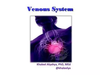
Function • Transport blood back to the heart • Prevent intravascular volume overload
The superficial , deep , perforating , and communicating veins The superficial venous system include the reticular veins as well as the great and the small saphenous veins and their tributaries. The reticular veins , a network of veins parallel to the skin surface and lying between the saphenous fascia and dermis, drain the lower extremity skin and subcutaneous tissue. The GSV usually lies directly on the muscular fascia in the saphenous compartment. The saphenous nerve lies anterior to the vein in the calf. A valve is present at the saphenofemoral junction in 94% to 100% of individuals. Approximately 60% of Small Saphenous Veins ( SSV ) join the popliteal vein within 8 cm of the knee joint, 20% join the GSV via anterior or posterior tributaries , and 20% join the femoral, or internal iliac veins. The sural nerve ascends immediately lateral to the vein. The deep venous system of the calf includes the tibial and peroneal veins as well as the soleal and gastrocnemial veins . 64 perforating veins between the ankle and the groin. The calf contain 4 groups of perforators ( medial : paratibial and posterior tibial veins ) , anterior and lateral perforators. Communicating veins connect veins within the same system.
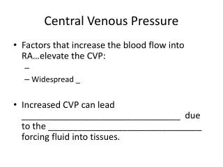
Valves • Venous valves: • One way • Two cusps of CT skeleton covered by endothelium • Closure at > 30cm/s • Exception: IVC, common iliacs, portal, cranial sinus
Etiology • Pregnancy • Pelvic obstruction • Chronic straining • Prolonged standing • Prolonged sitting
Etiology • Wearing constricting clothing • Obesity • Hormones • Heredity risk? • Both parents = 80% • 50/50 chance if one parent • 20% chance if neither parent
SCLEROTHERAPY • Acts by destroying the venous endothelial cells, exposing the subendothelial collagen fibers, and ultimately, the formation of a fibrotic obstruction. • In the US, FDA approved agents for sclerotherapy include sodium tetradecyl sulphate (STS), polidocanol, sodium morrhuate, and glycerine which is usually used with epinephrine. • A recent RCT found no advantage to compression bandaging for > 24 hours when thromboembolus-deterrent stockings were worn for the remainder of 14 days. • Complications : most complications are minor, and include matting, pigmentation, pain, allergy, and skin urticaria, these minor complications can be observed in 30% of patients. Severe complications are very rare <0.01% ( death, anaphylaxis, pulmonary embolus and stroke)
OPEN VENOUS SURGERY Ligation and stripping of the GSV or SSV, combined with excision of large VV, has been the standard of care. Recognition of frequent saphenous nerve injury during ankle-to-groin stripping and a better understanding of the venous hemodynamics changed the technique to a limited, groin-to-knee stripping. TYPES: HIGH LIGATION, DIVISION, AND STRIPPING OF THE GSV OR SSV. MINIPHLEBECTOMY TO REMOVE THE BULGING VV THROUGH A SMALL STAB WOUNDS. CRYOSTRIPPING. POWERED PHLEBECTOMY (TIPP). CHIVA METHOD (reentry perforators).
ENDOVENOUS THERMAL ABLASION Is relatively new, minimally invasive percutaneous procedure with several advantages over standard open surgery. It includes EVLA and RFA; a third technique that recently emerged includes the use of superheated steam, which destroys the endothelial layer and causes shrinkage of the collagen. Duplex U/S recognition of all refluxing venous segments and their ablation during procedure is the key to minimizing recurrence of VV. Inappropriate vein size (<2mm and >15mm for RFA), history of superficial thrombophlebitis resulting in partially obstructed saphenous vein, and tortuous GSV by U/S are potential C/I; patients with ropy VV or those with aneurysmal dilatation of SFJ are better served with HL/S. relative C/I include uncorrectable coagulopathy, liver dysfunction, limited use of local anesthsia, immobility, pregnancy, and breast feeding. Postprocedural care : graduated compression stocking with ankle pressure of 30-40 mm Hg or an elastic or nonelastic wrap is placed on the leg at the end of the procedure, recent evidence suggest elastic compression for 1 week (day and night), we suggest duplex U/S 24- 72 hours postprocedural to exclude any thrombotic event (GRADE 2C). COMPLICATIONS: EVLA : in an international endovascular working group registry that included 3696 procedures (bruising was 75%, parasthesia was 3%, thrombophlebitis was 1.87%, skin burn was 0.46%, and DVT or heat –induced thrombosis in 0.27%0. RFA : serious complications such as DVT or thermal skin injury, were not observed in a multicentre, nonrandomized study .( Parasthesia was observed in 3.2%, thrombophlebitis in 0.8%, ecchymosis along the course of GSV in 6.3% and skin pigmentations in 2%).
CLASSIFICATION OF CHRONIC VENOUS DISORDERS: CEAP • ONE: CLINICAL CLASSIFICATION :
Classification: CEAP *Eklof et al. J of Vasc Surg 2004 Clinical classification • C0: no visible or palpable signs of venous disease • C1: telangiectasies or reticular veins • C2: varicose veins • C3: edema • C4a: pigmentation or eczema • C4b: lipodermatosclerosis or atrophie blanche • C5: healed venous ulcer • C6: active venous ulcer • S: symptomatic, including ache, pain, tightness, skin • irritation, heaviness, and muscle cramps, and other • complaints attributable to venous dysfunction • A: asymptomatic
Worn during the day Elastic stockings with adjustments in pressure Lower pressure stockings (20-30mm Hg) for edema and DVT prophylaxis Higher pressure (30-40+mm Hg) for ulcers and significant venous disease Operator dependent Difficult to put on Physical impediments/Co-morbidities 50% of patients were unable to them on alone 30-65% noncompliance noted in clinical trials in venous centers Compression Stockings
COMPRESSION TREATMENT It is recommended to decrease ambulatory venous hypertension to patients with CVD in addition to lifestyle modification. The different forms include elastic stockings , paste gauze boots (Unna boots), multilayer wraps…. Pressure to compress the superficial veins in supine position range from 20-25 mmHg. When upright, pressure of 35-40 mmHg have been shown to narrow the superficial veins, and pressure > 60 mmHg are needed to occlude them. Most class C1-C2 require pressure 10- 20 mmHg; class C3-C4 require pressure of 20- 30 mmHg; while class C5-C6 require pressure of 30-40 mmHg On the basis of high- quality clinical evidence, the Guideline Committee recommends compression therapy for patients with CVI ( class C3 –C6 ) including those with leg ulcers. Arterial occlusive disease is contraindation to conventional high- pressure compression treatment. This especially true for patients with ABI < 0.5, in this situation and in those with restricted walking ability, intermittent pneumatic compression pumps may be useful adjunct in ulcer healing. Pneumatic compression devices applied primarily at night, are also used in patients with refractory edema and venous ulcers. The exact mechanism is unknown. But high pressure bandages do reduce venous reflux and improve calf muscle pump function, and increase subcutaneous tissue pressure may decrease edema thereby improving local metabolism by enhancing oxygen and nutrient diffusion to the skin and subcutaneous tissue. Finally, cytokines, as VEGF and TNF-< have been demonstrated to decrease with compression therapy .
Deep Vein Thrombosis (DVT): Epidemiology • Major health issue in industrialized countries • ~ 200.000 new cases diagnosed each year • The annual incidence 0.1 percent in young aduts • 1 percent in adults > 60 years old • Consequences can be serious and chronic or fatal • Pulmonary embolism (major cause of sudden death) • Postthrombotic syndrome • Phlegmasia cerulea dolens Gray HW. Semin Nucl Med. 2002;32(3):159-172. Feied C, Handler J. eMedicine. 2002;3(1). Accessed August 27, 2002. Mewissen MW, Seabrook GR, Meissner MH, et al. Radiology. 1999;211(1):39-49. Meignan M, Rosso J, Gauthier H, et al. Arch Intern Med. 2000;160(2):159-164.
Pathophysiology Virchow’s Triad : 1856 Venous thrombosis Phlegmasia dolens Mondor’s disease Venous Insuffenciency Variceal hemorrhage Pulmonary thromboembolism Paradoxical embolism
Blood Clotting • Vascular Phase • Platelet Phase • Coagulation Phase • Fibrinolytic Phase
Vascular Phase • Vasoconstriction • Exposure to tissues activate Tissue factor and initiate coagulation Tissue Factor
Platelet phase • Non-nucleated - arise from magakaryocytes • blood vessel wall (endothelial cells) prevent platelet adhesion and aggregation • platelets contain receptors for fibrinogen and von Willebrand factor • after vessel injury Platelets adhere and aggregate. • Release permeability increasing factors (e.g. vascular permeability factor, VPF) • Loose their membrane and form a viscous plug
Coagulation Phase • Two major pathways • Intrinsic pathway • Extrinsic pathway • Both converge at a common point • 13 soluble factors are involved in clotting • Biosynthesis of these factors are dependent on Vitamin K1 and K2 • Most of these factors are proteases • Normally inactive and sequentially activated • Hereditary lack of clotting factors lead to hemophilia -A m
Risk Factors • AMI • Antithrombin III deficiency • Behcet’s disease • Blood type A • Burns • Catheters • Chemotherapy • Estrogen replacements • Fibrinogen abnormality • Fractures • Hemolytic anemias • Heparin-associated thrombocytopenia • Homocysteinuria • Hyperlipidemias Immobilization Inflammatory bowel disease Malignancy Obesity Old age Plasminogen activator abnormality Polycythemia Postoperative Pregnancy Protein C deficiency Protein S deficiency Superficial phlebitis Trauma Varicose veins Venous stasis Warfarin (first few days of therapy)
D-DIMER TESTS DOPPLER ULTRASONOGRAPHY HELICAL COMPUTED TOMOGRAPHY CONTRAST VENOGRAPHY IMPEDANCE PLETHYSMOGRAPHY EMERGING TECHNOLOGIES Diagnosis
Thrombosis of the vena cava. A, Direct imaging. B, Digitally subtracted imaging. A vena cava filter is present at the cephalic aspect of the thrombus, having captured mboli from the lower extremity.
Treatment of Venous Thrombosis and Pulmonary Embolism : Goals • Prevent death from PE • Prevent post-thrombotic characterized by chronic deep venous insufficiency, chronic pain, venous stasis, recurrent cellulitis, and lower extremity ulceration. • Prevent recurrent venous thromboembolism (VTE) • Achieve these objectives with minimal side effects and inconvenience
Treatment of VTE : modalities • Anticoagulants • Thrombolytic therapy • Caval interruption • Surgical removal
Anticoagulants • Initial treatment with heparin is necessary. • Induction period with heparin therapy can be reduced to 5 days. • Treatment following hospital discharge is necessary. • LMWH is a major advance. • Optimal therapeutic range with warfarin established. • Optimal duration of warfarin therapy still to be established. • New oral small-molecule direct thrombin inhibitor.*
Thrombolytic Therapy • Not often indicated in venous thrombosis • Useful in major PE • Possible new indication in PE
Caval Filter • A single randomized trial has defined advantages and drawbacks of caval filters. The results indicate that inferior venacaval filters: a) Prevent recurrent PE in short term b) Increase the risk of recurrent deep venous thrombosis (DVT) in the long term
Anticoagulants • Heparin • Vitamin K antagonists (warfarin) • LMWH • Danaparoid* • Hirudin* • Pentasaccharide* • Oral small-molecule direct thrombin inhibitor* *Danaparoid is not approved by the FDA for use in the treatment of thrombosis or HIT. Natural hirudin is not approved by the FDA for any indication; recombinant hirudin (lepirudin) is approved for the treatment of thrombosis associated with HIT. Pentasaccharide and the new oral small-molecule direct thrombin inhibitor do not have FDA approval for any indication.
Anticoagulants Heparin Injection LMWH Injection Warfarin Oral Danaparoid* Injection Hirudin* Injection Pentasaccharide* Injection: Phase 3 Small molecule DTI†* Oral: Multiple clinical trials
Established Guidelines • Initial treatment with heparin is necessary. • Induction period with heparin therapy can be reduced to 5 days. • LMWH can replace heparin and is now treatment of choice. • Continued treatment following hospital discharge is necessary. • Optimal therapeutic range with warfarin is an INR of 2.0 to 3.0. • Optimal duration of warfarin therapy still to be established.
Unfractionated Heparin (UFH) • Sulphated carbohydrate • Purified from bovine lungs • Active in vitro and in vivo • Binds to a variety of cells and plasma proteins, leading to unpredictable effects • Short half-life : 1 - 5 hrs - monitor aPTT • Increased risk of heparin-induced thrombocytopenia (HIT) • Administration - parenteral- Do not inject IM - only IV or deep s.c. • Adverse effect - hemorrhage - antidote - protamine sulphate