Download
1 / 3
30 likes | 174 Views
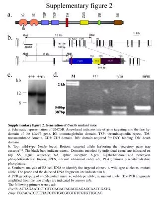
Guillemin et al. Supplementary figure 2. Supplementary figure 2 . Expression of BCL2L10 in human oocytes analyzed by immunofluorescence.

E N D
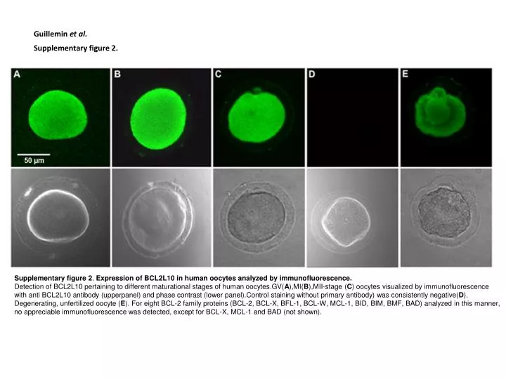
Guillemin et al. Supplementary figure 2. Supplementary figure 2. Expression of BCL2L10 in human oocytes analyzed by immunofluorescence. Detection of BCL2L10 pertaining to different maturational stages of human oocytes.GV(A),MI(B),MII-stage (C) oocytes visualized by immunofluorescence with anti BCL2L10 antibody (upperpanel) and phase contrast (lower panel).Control staining without primary antibody) was consistently negative(D). Degenerating, unfertilized oocyte (E). For eight BCL-2 family proteins (BCL-2, BCL-X, BFL-1, BCL-W, MCL-1, BID, BIM, BMF, BAD) analyzed in this manner, no appreciable immunofluorescence was detected, except for BCL-X, MCL-1 and BAD (not shown).
Supplementary figure 3. Supplementary figure 3. (A) Yeast two-hybrid interaction matrix for BCL2L10, BCL2, BAX and binding partners isolated in the screen. Yeast clones expressing BCL2L10, BCL2, and BAX in bait vector pGILDA were mated by replica plating with clones expressing BCL2L10, BAX, p53, ß-actin, -actin, HINT1 and TCTP in prey vector pJG4-5. Picture was taken after 48 h incubation at 30°C. Interactions are measured by the development of a blue color following induction of the LacZ reporter gene. BCL2L10 appears to bind to BAX, HINT1and TCTP as revealed by light blue coloration. BCL2 binds most strongly to BAX and TCTP, as well as to ß-actin, -actin and HINT1. BAX binds to itself but not to the other proteins. Expression of the different proteins has been checked by Western blotting (notshown). The BAX-BAX and BAX-BCL2 interactions served as positive controls for the yeast two-hybrid interaction assay. (B) Histidin pull down assay of the interaction in vitrobetween GST-BCL2L10 and TCTP-His6.TCTP-His6was fixed on Ni-NTA beads and GST (lane 2) or GST-BCL2L10 (lane 3) was applied on the complex. After elution, the attached protein complexes were resolved in a 12% polyacrylamidegel. Anti-GST and anti-TCTP antibodies were used in the Western blot. TCTP-His6bound specifically to GST-BCL2L10 (lane 3), whereas no interaction between TCTP-His6and GST was detected (lane2). Lane 1 (control): GST-BCL2L10 did not interact with Ni-NTA resin. (C) Indirect ELISA binding assay. Increasing concentrations of purified His-tagged TCTP was used to coat wells of a 96-well plate. Serial dilutions of GST-BCL2L10 were incubated with immobilized TCTP-His6. Binding was detected using anti-GST antibody. GST-BCL2L10 demonstrates dose-dependent binding to immobilized TCTP-His6. GST alone did not demonstrate significant binding to immobilized TCTP-His6. Intensity of the linear signal obtained with GST was subtracted from each experimental point.
Guillemin et al. Supplementary figure 4. Supplementary figure 4. Confocal images of in vitro matured human oocytes showing TCTP/tubulin double-staining. MII-stage human oocytes (C, D) were double-stained with antibodies against TCTP and microtubules. Panels A5-A8 and B5-B8, are enlarged images from A1-A4 and B1-B4, respectively. Panel A and panel B represent two different focal planes of the same oocyte.Microtubules and TCTP are abundant in the peripheral region (A4, A8) and around the meiotic apparatus (B4, B8). Scale bar, 10μm.