Download
1 / 1
10 likes | 78 Views
Enhancement of colonization on mouse spermatogonial stem cell by Low intensity ultrasound stimulation Mohaqiq M 1* , Movahedin M 1 , Mokhtari M 2 , Mazaheri Z 1 1: Anatomical Sciences Department, Faculty of Medicine, Tarbiat Modares University, Tehran, Iran.

E N D
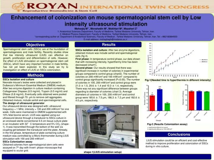
Enhancement of colonization on mouse spermatogonial stem cell by Low intensity ultrasound stimulation Mohaqiq M1*, Movahedin M1, Mokhtari M2, Mazaheri Z1 1: Anatomical Sciences Department, Faculty of Medicine, Tarbiat Modares University, Tehran, Iran. 2: Medical Physics Department, Faculty of Medicine, Tarbiat Modares University, Tehran, Iran. *corresponding author at: Department of Anatomical Sciences, Faculty of Medical , Tarbiat Modares University, Tehran, Iran. Tel: +982182884503 E-mail address: mohaghegh@modares.ac.ir Results Objectives Spermatogonial stem cells (SSCs) are at the foundation of spermatogenesis and male fertility. Recently studies show that low intensity ultrasound (LIUS) can effective on growth, proliferation and differentiation of cells. However, the effect of LIUS stimulation on spermatogonial stem cell (SSCs), which have very important function in male fertility, has not yet been explored. In this study we try to investigation on effect of LIUS on SSCs colonization. SSCs isolation and culture: After two enzyme digestions, obtained mixture was include sertoli and spermatogonial cells (fig 4). First phase: In temperature control phase, our data shown that with increasing intensity, hyperthermy time has been decreased (chart1). Second phase: Our results showed that there was significant increase in number of colonies in experimental groups compared to control group (chart2). The number of colonies on 200 mW/cm2 and 100 mW/cm2 compared to control group significantly increased (P<0.05). The number of colonies in 100, 200 mW/cm2 and control group were 21.0 ± 1.5, 29.6 ± 1.4 and 10.3 ± 1.4, respectively. There was not any significant difference between groups regarding to diameter of colonies (chart 3). Average diameters of colonies in 100, 200 mW/cm2 and control group were 185.6 ± 7.5 µm, 186.3 ± 7.2 µm and 182.6 ± 4.0 µm, respectively. Methods SSCs Isolation and culture Neonate mouse’s testes were collected and placed in Dulbecco’s Minimum Essential Medium (DMEM) medium. After two enzyme digestion in culture medium containing Collagenase/ Dispase (0.5 mg/ml), Trypsin (0.5 mg/ml) and DNase (0.8 mg/ml), Eventually cells obtained were pooled and filtered through 70- µm to remove cell aggregates. Obtained mixture, include sertoli and spermatogonial cells. The design of ultrasound generator Our ultrasound device was designed with: ultrasound frequency = 1 MHz, intensity = 100 and 200 mW/cm2 for one week. Cells were maintained in DMEM supplemented with 10% fetal bovine serum. LIUS was applied using our ultrasound device through a transducer to SSCs culture in an enclosed sterile conventional 3.5 cm tissue culture plate in an incubator with 32°C temperature and 5% CO2 (shape-1). It was transmitted through the bottom of the well via coupling gel between the transducer and the plate. Already, in the first phase, temperature of plate containing culture medium was controlled by a Micro thermometer during LIUS stimulation with 100 and 200 mW/cm2 . SSCs Colonization assay Obtained colonies from spermatogonial stem cells were assayed on 7th day with Invert- phase microscope that equipped to ocular grid. Fig-1(Needed time to hyperthermia in different intensity) Fig-2 (Results Colonization assay) References a: significantly different to control b: significantly different to 100 mW/cm2 Conclusions LIUS stimulation could be an efficient and cost-effective method to improve proliferation and colonization of SSCs during in vitro culture. shape-1(LIUS stimulation setup)