Download

1 / 44
570 likes | 1.94k Views
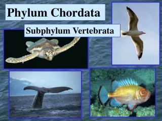
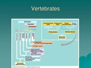
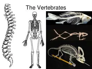
Vertebrates (subphylum vertebrata). Possess a backbone (aka vertebral column, spine) Vertebrae=Dorsal row of hollow skeletal elements (usually bone) Nerve cord=spinal cord, protected by vertebrae, (part of nervous system), ends in brain Bilateral symmetry, endoskeleton.

E N D
Vertebrates (subphylum vertebrata) • Possess a backbone (aka vertebral column, spine) • Vertebrae=Dorsal row of hollow skeletal elements (usually bone) • Nerve cord=spinal cord, protected by vertebrae, (part of nervous system), ends in brain • Bilateral symmetry, endoskeleton
Fish Form & FunctionGoals for this lab • Learn about fish: Topics • Skin/scales • Coloration • Locomotion • Fins • Muscles • Discuss 3 classes of fish • Dissect different fish- up to 3 different forms • Write paper comparing different fish forms • Due next Monday/Tuesday • Details to follow
Global Habitats 41.2% 58.2% 39.9%
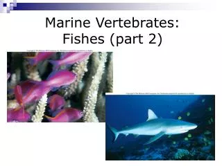
Fish importance • Appeared > 500 mya • Comprise half of vertebrate species • Feed on all types of marine organisms • some organisms previously discussed use fish as their home (bacteria to crustaceans) • Some animals eat fish • Most economically important marine organism • Vital source of protein to millions of humans • Ground up for chicken feed, fertilizer, leather, glue, vitamins obtained from them • Some kept as pets
Fish Morphology • Skin • Color • Bioluminescence • Swimming Locomotion • Fins • Muscles
Skin • Organ of the body • Consists of connective tissue • Muscles pull against skin tissue & skeleton • Key component of the muscle-tendon-tail fin system • Layers • Epidermis • Typically 250 m thick 10-30 cell layers • Range 20 m – 3 mm • Dermis
Fish Skin • Function: • Hold fish together • Serves as barrier against abrasive agents • Osmoregulation (what does this mean?) • Permeable respiratory function • Biomechanical properties in sharks
Fish Skin Derivatives: • Mucous formed in epidermis cells • Protect against infection • Constantly shed to remove bacteria and fungus • Ex. Clingfish lack scales, protect their bodies by a thick layer of mucous • Bone is also skin derivative • scales, most important
Fish Scales • First appear as dermal bone • Found in fossil of Cambrian period (570 mya) • Layered bone, solid armor-constrained movement • Evolved smaller and reduced into scales • 5 types of scales (examples with images to follow) • Placoid • Cosmoid • Ganoid • Cycloid • Ctenoid
Fish Scales: Placoid • Found in elasmobranchs (sharks & rays) • “teeth like”, same composition • As fish grows, do not increase in size, instead new scales are added
Fish Scales: Cosmoid • In the Sarcopterygii (fish with fleshy lobe fins), primitive fish • Less evolved than Elasmobranchs and Actinopterygii (fish with rayed fins) • Scales found in fossil record but not in any living fish, • Except in simplified version of coelocanth and lungfish
Fish Scales: Ganoid • In primitive Actinopterygii • Found in reedfish, polypterus, gar, bowfin, and sturgeons • Were thick heavy scales when first appeared • Rhomboid-shaped • Developed into teleost scales
Ctenoid scales Cycloid scales Fish Scales: Teleost scales • Two types: • Ctenoid-higher fish • Cycloid-soft-rayed, anchovies, sardine • Mineralized surface layer & inner collagenous layer • Scales surrounded by dermis, in dermal pockets • Grow from top, bottom, and insides; overlap lower part • Scales grow with fish • Characterized by concentric ridges (growth increments)
Coloration • Fish display a multitude of patterns involving • 2 or more colors, • in many tints and shades, • arranged in spots, stripes, patches, and blotches • 3 Types of coloration predominant in oceans • Silver – pelagic, upper zone • Red – deeper zone (~ 500 m) • Black or violet – deep sea • Countershaded near shore and colorful in coral reefs
Coloration • Chromatophores • Colored cells from which light is reflected off • Located in the skin (dermis), eyes • Various colors/hues-combination of different chromatophores • Functional Roles of Colors in Fishes-examples of each to follow • Social Roles • Advertisement • Mimicry • Hiding • Protection from sun (especially larvae)
Coloration: Social roles Cleaner Fish:distinctive markings recognized by larger fish
Coloration: Advertisement: Bright, bold and showy males indicate: Reproductive availability, either permanently or seasonally, e.g. cichlids, wrasses, minnows, sunfish Unpalatable or venomous, e.g. lionfishes Mimicry – Disguise: Disguises: look like something in habitat, e.g. leaffish, sargasso fish Mimicry: mimic distasteful species
Coloration: Concealment General color resemblance – resemble background Variable color resemblance – change with background, e.g. flatfish Obliterative shading – countershading, dark above, light below (invisible fish) Disruptive coloration – disruptive contours that breakup outline; bold stripes, bars, false eye spots Coincident disruptive coloration – joining together of unrelated parts of the body to reduce recognition; e.g. sea dragon
Bioluminescence • Most luminous fish found 300-1000 m depths, few shallow • 3 Types of light producing methods: • Self-luminous (on/off) • Symbiotic bacteria nurtured in special glands • Acquire from other bioluminescent organisms- diet contains light-emitting compounds • Function: • Concealment by counter-illumination - ventral placement matches background from above, against attack from below • Dorsal photophores safeguard against predators from above • Advertisement for courting, maintaining territory, to startle and confuse predators, and feeding
Fish Locomotion Means of Locomotion: • Simplest form: Passive drifting of larval fish • Many can: • Burrow • Walk, hop, or crawl • Glide • Fly • Most can: • Swim in a variety of ways
Types of fins: • Paired fins: pectoral and pelvic • Median fins:dorsal, caudal, anal, & adipose Fins
Fins • Main functions: • Swimming – increase surface area w/o increasing mass • Stabilizers – yaw, stability-dorsal and anal fins • - brake, pitch, roll, reverse -pectoral/pelvic • thrust with caudal fin • Modifications in fins: • Defense – spines, enlarge fish • Locomotion – modified for crawling, flying, gliding • Hunting – lures, sensory organs • Respiratory organ – lungfish, supply oxygen to eggs
Fins Soft rays vs. Spines • Spines: • Usually hard and pointed • Unsegmented • Unbranched • Solid • Soft rays: • Usually soft and not pointed • Segmented • Usually branched • Bilateral, w/left and right halves
Fish Muscles • Muscles provide power for swimming • Myomers=bands of muscle, run along sides of body, attached to backbone • Constitute up to 80% of the fish itself • Much hardly used except during emergencies • Don’t have to contend with same effect of gravity • Fish muscle arrangement not suitable on land • Cow: 30% muscle/wt • Tuna: 60% muscle/wt • Contraction causes oscillation of body and tail • Body bends as one side contracts b/c of an incompressible • notochord or vertebral column • Caused by bands of muscle = myomeres
Fish Muscles • Major fibers (see handout): • Red, pink, and white • Pink intermediate between red and white • Muscle types do not intermingle • Different motor systems used for different swimming conditions • Red – cruising • White – short duration, burst swimming • Pink – sustained swimming, used after red and before white
Fish-Body shapes-see textbook for images (Figure 8.9) • Fusiform-spindle shaped, e.g. tuna • Compressiform-laterally compressed, angelfish, butterfly fish • Anguilliform-eel-like • Filiform-even smaller anguilliform, e.g. snipe eel
Body shapes continued • Depressiform-flatfish, rays, flounder • Taeniform-gunnel • Sagittiform-e.g. pike • Globiform-e.g. lumpsucker
Fish Locomotion Swimming classified into 2 generic categories: Periodic (or steady or sustained)- e.g. running marathons, for covering large distance at constant speed Transient (or unsteady) – e.g. like running sprints, used for catching prey or avoiding predators
Isolate and move only fin(s) pectoral Rajiform - pectoral Labriform -pectoral oscillate Diodontiform - pectoral anal Gymnotiform -anal dorsal Tetraodontiform – anal+dorsal Balistiform – anal+dorsal Amiiform -dorsal Ostraciform-rigid body, caudal main propulsion Flex caudal portion, fast swimmers Thunniform-rigid body, caudal main propulsion Carangiform Subcarangiform Undulate the body: eels, elongate fish Anguilliform (Wavelike) (fanlike)
http://www.oceanfootage.com/stockfootage/Titan_Trigger_Fish//?DVfSESSCKIE=7305db92882366fd26c463edc209393f8e25bdc9http://www.oceanfootage.com/stockfootage/Titan_Trigger_Fish//?DVfSESSCKIE=7305db92882366fd26c463edc209393f8e25bdc9
Tuna: Ultimate Living Swimming MachineSwim continuously – feeding, courtship, rest, reproduction
Tuna: Ultimate Living Swimming Machinehydrodynamic adaptations • Big size-high performance engine • Streamlining-spindle shaped & rigid body • Small structures at various parts of the body to improve swimming efficiency and reduce drag, e.g. • Eyes flush with body – don’t protrude • Adipose eyelid - smooth, reduce drag • Depression grooves for dorsal, pelvic, & pectoral fins at high speed • Keeled peduncle - cutting through water • Finlets for cross-flow - delayed separation
Tuna: Ultimate Living Swimming Machine • Must swim to survive: • No gas bladder, rigid body, ram ventilation • High blood volume, large heart, maintain warm core (25oC) • School to utilize vortices generated by other fish (~like race car driver who “slipstreams” and then slingshots past leading car) • Adopt swim-glide for energy savings (like birds) • High narrow tails – propulsion with least effort, used to design efficient propulsion systems for ships Slipstream: The area of reduced pressure or forward suction produced by and immediately behind a fast-moving object as it moves through air or water.
Fish-mouth types (some) • Large mouth with teeth (e.g. barracuda) • Long snout/small mouth (e.g. butterfly fish) • Protrusible mouth (e.g. slipmouth) • Beak-like mouth (e.g. parrotfish) • Large mouth (e.g. herrings)
Fish Three Classes: Agnatha Chondrithyes Osteicthyes
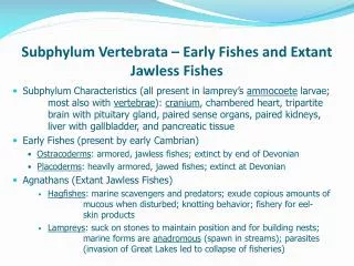
Class Agnatha • Jawless fishes • Ex. Hagfish, lampreys • No paired fins • Gill holes, no slits or operculum • Large sucking mouth with teeth • Scavengers • As a defense mechanism, secrete slime then tie itself in knots to escape predators • Also tie in knots for pulling food off carcasses, and cleaning slime from body
Class Agnatha Hagfish’s mouth http://www.soest.hawaii.edu/oceanography/faculty/csmith/index.html
Class Chondricthyes • Sharks and rays • Skeleton = cartilage, not bone • Paired fins-efficient swimming • Gill slits exposed, • no operculum • Large oil-filled liver • Heterocercal tail (upper longer than lower lobe) • Placoid scales-skin like sandpaper
Class Osteichthyes • Bony fish • Largest group of living vertebrates • Bones for skeletons • Gill covering (operculum) • Swim bladder (balloon-like) • Homocercal tails (even) • Cycloid & Ctenoid scales
Dissection Worksheet • Working in groups of 2 or 3 people, • dissect 1 fish following the worksheet and writing the answers to the questions in your notebook as you go. • Need to draw 3 external illustrations in your notebooks • 1 of the fish you are dissecting, before you dissect it • 2 others that have specialized mouths and caudal fins • label the type of mouth and caudal fin each has • Label the following structures on each illustration: • gill cover, pectoral fins, pelvic fins, dorsal fin, • anal fin, adipose fin (if present), lateral line • give the head length, total length, and the fork length (of the dissected one ONLY, see handout) • look at a scale under a microscope and draw it.
Dissection Worksheet continued • Cut through body cavity • Find the following • Heart • Liver • Stomach/intestines • Swim bladder (if applicable) • Spine • Cut cross section, 2/3 down the body • Red muscle • White muscle
Scales- use slides • Draw • Placoid • Ganoid • Cycloid • Ctenoid