Download
1 / 45
450 likes | 541 Views
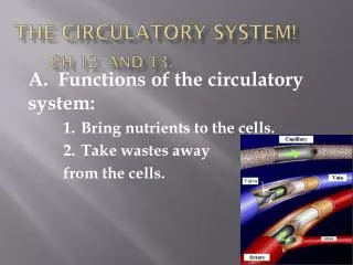
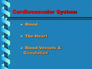
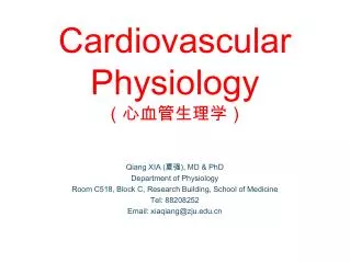
The Cardiovascular System: Blood and Sheep’s Heart. Blood. Liquid connective tissue 3 general functions Transportation Gases, nutrients, hormones, waste products Regulation pH, body temperature, osmotic pressure Protection Clotting, white blood cells, proteins.

E N D
Blood • Liquid connective tissue • 3 general functions • Transportation • Gases, nutrients, hormones, waste products • Regulation • pH, body temperature, osmotic pressure • Protection • Clotting, white blood cells, proteins
Components of Blood • Blood plasma – water liquid extracellular matrix • 91.5% water, 8.5% solutes (primarily proteins) • Hepatocytes synthesize most plasma proteins • Albumins, fibrinogen, antibodies • Other solutes include electrolytes, nutrients, enzymes, hormones, gases and waste products • Formed elements – cells and cell fragments • Red blood cells (RBCs) • White blood cells (WBCs) • Platelets
Formation of Blood Cells • Negative feedback systems regulate the total number of RBCs and platelets in circulation • Abundance of WBC types based of response to invading pathogens or foreign antigens • Hemopoiesis or hemotopoiesis • Red bone marrow primary site • Pluripotent stem cells have the ability to develop into many different types of cells
Formation of Blood Cells • Stem cells in bone marrow • Reproduce themselves • Proliferate and differentiate • Cells enter blood stream through sinusoids • Formed elements do not divide once they leave red bone marrow • Exception is lymphocytes
Formation of Blood Cells • Pluripotent stem cells produce • Myeloid stem cells • Give rise to red blood cells, platelets, monocytes, neutrophils, eosinophils and basophils • Lymphoid stem cells give rise to • Lymphocytes • Hemopoietic growth factors regulate differentiation and proliferation • Erythropoietin – RBCs • Thrombopoietin – platelets • Colony-stimulating factors (CSFs) and interleukins – WBCs
Red Blood Cells/ Erythrocytes • Contain oxygen-carrying protein hemoglobin • Production = destruction with at least 2 million new RBCs per second • Biconcave disc – increases surface area • Strong, flexible plasma membrane • Glycolipids in plasma membrane responsible for ABO and Rh blood groups • Lack nucleus and other organelles • No mitochondria – doesn’t use oxygen
Hemoglobin • Globin – 4 polypeptide chains • Heme in each of 4 chains • Iron ion can combine reversibly with one oxygen molecule • Also transports 23% of total carbon dioxide • Combines with amino acids of globin • Nitric oxide (NO) binds to hemoglobin • Releases NO causing vasodilation to improve blood flow and oxygen delivery
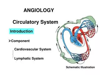
Red Blood Cells • RBC life cycle • Live only about 120 days • Cannot synthesize new components – no nucleus • Ruptured red blood cells removed from circulation and destroyed by fixed phagocytic macrophages in spleen and liver • Breakdown products recycled • Globin’s amino acids reused • Iron reused • Non-iron heme ends as yellow pigment urobilin in urine or brown pigment stercobilin in feces
Circulation for about 120 days Circulation for about 120 days Circulation for about 120 days Circulation for about 120 days Circulation for about 120 days Circulation for about 120 days 7 7 7 7 7 7 7 3 3 3 3 3 3 3 3 3 3 3 Reused for protein synthesis Reused for protein synthesis Reused for protein synthesis Reused for protein synthesis Reused for protein synthesis Reused for protein synthesis Reused for protein synthesis Reused for protein synthesis Reused for protein synthesis Reused for protein synthesis Reused for protein synthesis Amino acids Amino acids Amino acids Amino acids Amino acids Amino acids Amino acids Amino acids Amino acids Amino acids Amino acids Transferrin Transferrin Transferrin Transferrin Transferrin Transferrin Transferrin Transferrin Fe3+ Fe3+ Fe3+ Fe3+ Fe3+ Fe3+ Fe3+ Fe3+ Globin Globin Globin Globin Globin Globin Globin Globin Globin Globin Globin Globin 6 6 6 6 6 6 6 6 4 4 4 4 4 4 4 4 4 4 5 5 5 5 5 5 5 5 5 Fe3+ Fe3+ Fe3+ Fe3+ Fe3+ Fe3+ Fe3+ Fe3+ Fe3+ Fe3+ 2 2 2 2 2 2 2 2 2 2 2 2 Fe3+ Fe3+ Fe3+ Fe3+ Fe3+ Fe3+ Fe3+ Ferritin Ferritin Ferritin Ferritin Ferritin Ferritin Ferritin Ferritin Ferritin Heme Heme Heme Heme Heme Heme Heme Heme Heme Heme Heme Heme Transferrin Transferrin Transferrin Transferrin Transferrin Transferrin Transferrin Transferrin Transferrin Transferrin + + + + + + + Globin Globin Globin Globin Globin Globin Globin Bilirubin Bilirubin Bilirubin Bilirubin 9 9 9 9 9 + + + + + + + Biliverdin Biliverdin Biliverdin Biliverdin Biliverdin Liver Liver Liver Liver Liver Liver Liver Liver Liver Vitamin B12 Vitamin B12 Vitamin B12 Vitamin B12 Vitamin B12 Vitamin B12 Vitamin B12 Bilirubin Bilirubin Bilirubin Bilirubin Bilirubin 11 11 11 1 1 1 1 1 1 1 1 1 1 1 1 1 Red blood cell death and phagocytosis Red blood cell death and phagocytosis Red blood cell death and phagocytosis Red blood cell death and phagocytosis Red blood cell death and phagocytosis Red blood cell death and phagocytosis Red blood cell death and phagocytosis Red blood cell death and phagocytosis Red blood cell death and phagocytosis Red blood cell death and phagocytosis Red blood cell death and phagocytosis Red blood cell death and phagocytosis Red blood cell death and phagocytosis + + + + + + + 10 10 10 10 Erythopoietin Erythopoietin Erythopoietin Erythopoietin Erythopoietin Erythopoietin Erythopoietin Small intestine Small intestine Small intestine Kidney Kidney 8 8 8 8 8 8 Erythropoiesis in red bone marrow Erythropoiesis in red bone marrow Erythropoiesis in red bone marrow Erythropoiesis in red bone marrow Erythropoiesis in red bone marrow Erythropoiesis in red bone marrow Bilirubin Bilirubin Bilirubin 13 13 12 12 12 Urobilin Urobilin Urobilinogen Urobilinogen Urobilinogen Macrophage in spleen, liver, or red bone marrow Macrophage in spleen, liver, or red bone marrow Macrophage in spleen, liver, or red bone marrow Macrophage in spleen, liver, or red bone marrow Macrophage in spleen, liver, or red bone marrow Macrophage in spleen, liver, or red bone marrow Macrophage in spleen, liver, or red bone marrow Macrophage in spleen, liver, or red bone marrow Macrophage in spleen, liver, or red bone marrow Macrophage in spleen, liver, or red bone marrow Macrophage in spleen, liver, or red bone marrow Macrophage in spleen, liver, or red bone marrow Macrophage in spleen, liver, or red bone marrow Key: Key: Key: Key: Key: Key: Key: Key: Key: Key: Key: Key: Key: Bacteria Bacteria Bacteria in blood in blood in blood in blood in blood in blood in blood in blood in blood in blood in blood in blood in blood Stercobilin Stercobilin Stercobilin 14 Large intestine in bile in bile in bile in bile in bile in bile in bile in bile in bile in bile in bile in bile in bile Feces Feces Feces Urine Urine
Starts in red bone marrow with proerythroblast Cell near the end of development ejects nucleus and becomes a reticulocyte Develop into mature RBC within 1-2 days Negative feedback balances production with destruction Controlled condition is amount of oxygen delivery to tissues Hypoxia stimulates release of erythropoietin Erythropoiesis
White Blood Cells/ Leukocytes • Have nuclei • Do not contain hemoglobin • Granular or agranular based on staining highlighting large conspicuous granules • Granular leukocytes • Neutrophils, eosinophils, basophils • Agranular leukocytes • Lymphocytes and monocytes
Functions of WBCs • Usually live a few days • Except for lymphocytes – live for months or years • Far less numerous than RBCs • Leukocytosis is a normal protective response to invaders, strenuous exercise, anesthesia and surgery • Leukopenia is never beneficial • General function to combat invaders by phagocytosis or immune responses
Many WBCs leave the bloodstream Emigration (formerly diapedesis) Roll along endothelium Stick to and then squeeze between endothelial cells Precise signals vary for different types of WBCs Emigration of WBCs
WBCs • Neutrophils and macrophages are active phagocytes • Attracted by chemotaxis • Neutrophils respond most quickly to tissue damage by bacteria • Uses lysozymes, strong oxidants, defensins • Monocytes take longer to arrive but arrive in larger numbers and destroy more microbes • Enlarge and differentiate into macrophages
WBCs • Basophila leave capillaries and release granules containing heparin, histamine and serotonin, at sites of inflammation • Intensify inflammatory reaction • Involved in hypersensitivity reactions (allergies) • Eosinophils leave capillaries and enter tissue fluid • Release histaminase, phagocytize antigen-antibody complexes and effective against certain parasitic worms
Lymphocytes • Lymphocytes are the major soldiers of the immune system • B cells – destroying bacteria and inactivating their toxins • T cells – attack viruses, fungi, transplanted cells, cancer cells and some bacteria • Natural Killer (NK) cells – attack a wide variety of infectious microbes and certain tumor cells
Platelets/ Thrombocytes • Myeloid stem cells develop eventually into a megakaryocyte • Splinters into 2000-3000 fragments • Each fragment enclosed in a piece of plasma membrane • Disc-shaped with many vesicles but no nucleus • Help stop blood loss by forming platelet plug • Granules contain blood clot promoting chemicals • Short life span – 5-9 days
Stem cell transplants • Bone marrow transplant • Recipient's red bone marrow replaced entirely by healthy, noncancerous cells to establish normal blood cell counts • Takes 2-3 weeks to begin producing enough WBCs to fight off infections • Graft-versus-host-disease – transplanted red bone marrow may produce T cells that attack host tissues • Cord-blood transplant • Stem cells obtained from umbilical cord shortly before birth • Easily collected and can be stored indefinitely • Less likely to cause graft-versus-host-disease
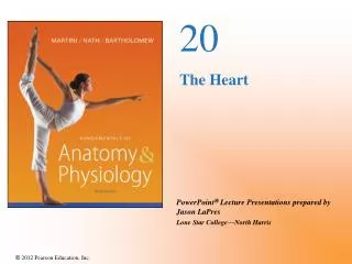
Hemostasis • Sequence of responses that stops bleeding • 3 mechanisms reduce blood loss • Vascular spasm • Smooth muscle in artery or arteriole walls contracts • Platelet plug formation • Platelets stick to parts of damaged blood vessel, become activated and accumulate large numbers • Blood clotting (coagulation)
Red blood cell Red blood cell Red blood cell Platelet Platelet Platelet Collagen fibers and damaged endothelium Collagen fibers and damaged endothelium Collagen fibers and damaged endothelium 1 1 1 Platelet adhesion Platelet adhesion Platelet adhesion 1 1 1 Liberated ADP, serotonin, and thromboxane A2 Liberated ADP, serotonin, and thromboxane A2 2 2 2 2 Platelet release reaction Platelet release reaction Platelet plug 3 Platelet aggregation 3
Blood clotting Serum is blood plasma minus clotting proteins Clotting – series of chemical reactions culminating in formation of fibrin threads Clotting (coagulation) factors – Ca2+, several inactive enzymes, various molecules associated with platelets or released by damaged tissues Blood Clotting
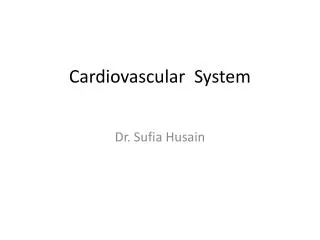
3 Stages of Clotting • Extrinsic or intrinsic pathways lead to formation of prothrombinase • Prothrombinase converts prothrombin into thrombin • Thrombin converts fibrinogen (soluble) into fibrin (insoluble) forming the threads of the clot
(a) Extrinsic pathway (a) Extrinsic pathway (a) Extrinsic pathway (b) Intrinsic pathway (b) Intrinsic pathway (b) Intrinsic pathway Tissue trauma Tissue trauma Tissue trauma Blood trauma Blood trauma Blood trauma Damaged endothelial cells expose collagen fibers Damaged endothelial cells expose collagen fibers Damaged endothelial cells expose collagen fibers Tissue factor (TF) Tissue factor (TF) Tissue factor (TF) Damaged platelets Damaged platelets Damaged platelets Activated XII Activated XII Activated XII Activated platelets Activated platelets Activated platelets Ca2+ Ca2+ Ca2+ Ca2+ Ca2+ Ca2+ + + Platelet phospholipids Platelet phospholipids Platelet phospholipids Activated X Activated X Activated X Activated X Activated X Activated X V V V V V V + + Ca2+ Ca2+ Ca2+ Ca2+ Ca2+ Ca2+ 1 1 1 PROTHROMBINASE PROTHROMBINASE PROTHROMBINASE (c) Common pathway (c) Common pathway Ca2+ Ca2+ Prothrombin (II) Prothrombin (II) 2 2 THROMBIN THROMBIN Ca2+ XIII Fibrinogen (I) Activated XIII STRENGTHENED Loose fibrin threads 3 FIBRIN THREADS
Blood Clotting • Extrinsic pathway • Fewer steps then intrinsic and occurs rapidly • Tissue factor (TF) or thromboplastin leaks into the blood from cells outside (extrinsic to) blood vessels and initiates formation of prothrombinase • Intrinsic pathway • More complex and slower than extrinsic • Activators are either in direct contact with blood or contained within (intrinsic to) the blood • Outside tissue damage not needed • Also forms prothrombinase
Blood Clotting: Common pathway • Marked by formation of prothrombinase • Prothrombinase with Ca2+ catalyzes conversion of prothrombin to thrombin • Thrombin with Ca2+ converts soluble fibrinogen into insoluble fibrin • Thrombin has 2 positive feedback effects • Accelerates formation of prothrombinase • Thrombin activates platelets • Clot formation remains localized because fibrin absorbs thrombin and clotting factor concentrations are low
Blood Groups and Blood Types • Agglutinogens – surface of RBCs contain genetically determined assortment of antigens • Blood group – based on presence or absence of various antigens • At least 24 blood groups and more than 100 antigens • ABO and Rh
ABO Blood Group • Based on A and B antigens • Type A blood has only antigen A • Type B blood has only antigen B • Type AB blood has antigens A and B • Universal recipients – neither anti-A or anti-B antibodies • Type O blood has neither antigen • Universal donor • Reason for antibodies presence not clear
Rh blood group People whose RBCs have the Rh antigen are Rh+ People who lack the Rh antigen are Rh- Normally, blood plasma does not contain anti-RH antibodies Hemolytic disease of the newborn (HDN) – if blood from Rh+ fetus contacts Rh-mother during birth, anti-Rh antibodies made Affect is on second Rh+ baby Hemolytic Disease
Single drops of blood are mixed with different antisera Agglutination with an antisera indicates the presence of that antigen on the RBC Typing Blood
The pump Cat heart showing pericardium
Features to focus on Be able to trace blood flow through the heart starting with the Superior Vena Cava Aorta Superior Vena Cava Pulmonary Trunk Right Atrium Fossa ovalis Right Ventricle Tricuspid valve Chordae tendineae Trabeculae carneae Pulmonary semilunar valves Left Atrium Mitral (bicuspid) valve Left Ventricle Papilary muscle Interventricular septum Exterior features Auricle Apex Coronary arteries and veins Coronary sinus SA node