Download
1 / 175
1.81k likes | 2.18k Views
20 The Heart. An Introduction to the Cardiovascular System. The Pulmonary Circuit Carries blood to and from gas exchange surfaces of lungs The Systemic Circuit Carries blood to and from the body Blood alternates between pulmonary circuit and systemic circuit.

E N D
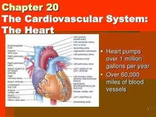
20 The Heart
An Introduction to the Cardiovascular System • The Pulmonary Circuit • Carries blood to and from gas exchange surfaces of lungs • The Systemic Circuit • Carries blood to and from the body • Blood alternates between pulmonary circuit and systemic circuit
An Introduction to the Cardiovascular System • Three Types of Blood Vessels • Arteries • Carry blood away from heart • Veins • Carry blood to heart • Capillaries • Networks between arteries and veins
An Introduction to the Cardiovascular System • Capillaries • Also called exchange vessels • Exchange materials between blood and tissues • Materials include dissolved gases, nutrients, waste products
Figure 20-1 An Overview of the Cardiovascular System PULMONARY CIRCUIT SYSTEMIC CIRCUIT Systemic arteries Pulmonary arteries Systemic veins Pulmonary veins Capillaries in head, neck, upper limbs Capillaries in lungs Left atrium Right atrium Left ventricle Right ventricle Capillaries in trunk and lower limbs
An Introduction to the Cardiovascular System • Four Chambers of the Heart • Right atrium • Collects blood from systemic circuit • Right ventricle • Pumps blood to pulmonary circuit • Left atrium • Collects blood from pulmonary circuit • Left ventricle • Pumps blood to systemic circuit
20-1 Anatomy of the Heart • The Heart • Great veins and arteries at the base • Pointed tip is apex • Surrounded by pericardial sac • Sits between two pleural cavities in the mediastinum
Figure 20-2a The Location of the Heart in the Thoracic Cavity Trachea Thyroid gland First rib (cut) Base of heart Left lung Right lung Apex of heart Diaphragm Parietal pericardium (cut) An anterior view of the chest, showing the position of the heart and major blood vessels relative to the ribs, lungs, and diaphragm.
20-1 Anatomy of the Heart • The Pericardium • Double lining of the pericardial cavity • Visceral pericardium • Inner layer of pericardium • Parietal pericardium • Outer layer • Forms inner layer of pericardial sac
20-1 Anatomy of the Heart • The Pericardium • Pericardial cavity • Is between parietal and visceral layers • Contains pericardial fluid • Pericardial sac • Fibrous tissue • Surrounds and stabilizes heart
Figure 20-2b The Location of the Heart in the Thoracic Cavity Posterior mediastinum Esophagus Aorta (arch segment removed) Left pulmonary artery Right pleural cavity Left pleural cavity Right lung Left lung Left pulmonary vein Bronchus of lung Pulmonary trunk Right pulmonary artery Aortic arch Left atrium Right pulmonary vein Left ventricle Pericardial cavity Superior vena cava Epicardium Right atrium Pericardial sac Right ventricle Anterior mediastinum A superior view of the organs in the mediastinum; portions of the lungs have been removed to reveal blood vessels and airways. The heart is situated in the anterior part of the mediastinum, immediately posterior to the sternum.
Figure 20-2c The Location of the Heart in the Thoracic Cavity Base of heart Wrist (corresponds to base of heart) Cut edge of parietal pericardium Inner wall (corresponds to epicardium) Fibrous tissue of pericardial sac Air space (corresponds to pericardial cavity) Parietal pericardium Areolar tissue Mesothelium Outer wall (corresponds to parietal pericardium) Cut edge of epicardium Balloon Fibrous attachment to diaphragm Apex of heart The relationship between the heart and the pericardial cavity; compare with the fist-and-balloon example.
20-1 Anatomy of the Heart • Superficial Anatomy of the Heart • Atria • Thin-walled • Expandable outer auricle (atrial appendage)
20-1 Anatomy of the Heart • Superficial Anatomy of the Heart • Sulci • Coronary sulcus divides atria and ventricles • Anterior interventricular sulcus and posterior interventricular sulcus • Separate left and right ventricles • Contain blood vessels of cardiac muscle
Figure 20-3a The Superficial Anatomy of the Heart Left common carotid artery Left subclavian artery Arch of aorta Brachiocephalic trunk Ligamentum arteriosum Descending aorta Ascending aorta Left pulmonary artery Superior vena cava Pulmonary trunk Auricle of right atrium Auricle of left atrium RIGHT ATRIUM Fat and vessels in anterior interventricular sulcus RIGHT VENTRICLE Fat and vessels in coronary sulcus LEFT VENTRICLE Major anatomical features on the anterior surface.
Figure 20-3a The Superficial Anatomy of the Heart Pulmonary trunk Auricle of left atrium Fibrous pericardium Ascending aorta Parietal pericardium Superior vena cava Auricle of right atrium RIGHT ATRIUM Right coronary artery Coronary sulcus RIGHT VENTRICLE Marginal branch of right coronary artery Parietal pericardium fused to diaphragm LEFT VENTRICLE Anterior interventricular sulcus Major anatomical features on the anterior surface.
Figure 20-3b The Superficial Anatomy of the Heart Arch of aorta Left pulmonary artery Right pulmonary artery Left pulmonary veins Fat and vessels in coronary sulcus Superior vena cava LEFT ATRIUM Coronary sinus Right pulmonary veins (superior and inferior) RIGHT ATRIUM LEFT VENTRICLE Inferior vena cava RIGHT VENTRICLE Fat and vessels in posterior interventricular sulcus Major landmarks on the posterior surface. Coronary arteries (which supply the heart itself) are shown in red; coronary veins are shown in blue.
Figure 20-3c The Superficial Anatomy of the Heart Base of heart 1 1 Ribs 2 2 3 3 4 4 Apex of heart 5 5 6 6 7 7 8 8 9 9 10 10 Heart position relative to the rib cage.
20-1 Anatomy of the Heart • The Heart Wall • Epicardium • Myocardium • Endocardium
20-1 Anatomy of the Heart Epicardium (Outer Layer) Visceral pericardium Covers the heart
20-1 Anatomy of the Heart • Myocardium (Middle Layer) • Muscular wall of the heart • Concentric layers of cardiac muscle tissue • Atrial myocardium wraps around great vessels • Two divisions of ventricular myocardium • Endocardium (Inner Layer) • Simple squamous epithelium
Figure 20-4a The Heart Wall Parietal pericardium Dense fibrous layer Areolar tissue Mesothelium Pericardial cavity Myocardium (cardiac muscle tissue) Epicardium (visceral pericardium) Cardiac muscle cells Mesothelium Connective tissues Areolar tissue Endocardium Areolar tissue Endothelium
Figure 20-4b The Heart Wall Atrial musculature Ventricular musculature Cardiac muscle tissue forms concentric layers that wrap around the atria or spiral within the walls of the ventricles.
20-1 Anatomy of the Heart • Cardiac Muscle Tissue • Intercalated discs • Interconnect cardiac muscle cells • Secured by desmosomes • Linked by gap junctions • Convey force of contraction • Propagate action potentials
Figure 20-5a Cardiac Muscle Cells Cardiac muscle cell Mitochondria Intercalated disc (sectioned) Nucleus Cardiac muscle cell (sectioned) Bundles of myofibrils Intercalated discs Cardiac muscle cells
Figure 20-5b Cardiac Muscle Cells Intercalated disc Gap junction Opposing plasma membranes Desmosomes Structure of an intercalated disc
Figure 20-5c Cardiac Muscle Cells Intercalated discs LM 575 Cardiac muscle tissue Cardiac muscle tissue
20-1 Anatomy of the Heart • Characteristics of Cardiac Muscle Cells • Small size • Single, central nucleus • Branching interconnections between cells • Intercalated discs
Table 20-1 Structural and Functional Differences between Cardiac Muscle Cells and Skeletal Muscle Fibers
20-1 Anatomy of the Heart • Internal Anatomy and Organization • Interatrial septum separates atria • Interventricular septum separates ventricles
20-1 Anatomy of the Heart • Internal Anatomy and Organization • Atrioventricular (AV) valves • Connect right atrium to right ventricle and left atrium to left ventricle • Are folds of fibrous tissue that extend into openings between atria and ventricles • Permit blood flow in one direction • From atria to ventricles
20-1 Anatomy of the Heart • The Right Atrium • Superior vena cava • Receives blood from head, neck, upper limbs, and chest • Inferior vena cava • Receives blood from trunk, viscera, and lower limbs • Coronary sinus • Cardiac veins return blood to coronary sinus • Coronary sinus opens into right atrium
20-1 Anatomy of the Heart • The Right Atrium • Foramen ovale • Before birth, is an opening through interatrial septum • Connects the two atria • Seals off at birth, forming fossa ovalis
20-1 Anatomy of the Heart • The Right Atrium • Pectinate muscles • Contain prominent muscular ridges • On anterior atrial wall and inner surfaces of right auricle
Figure 20-6a The Sectional Anatomy of the Heart Left common carotid artery Left subclavian artery Brachiocephalic trunk Ligamentum arteriosum Pulmonary trunk Superior vena cava Aortic arch Pulmonary valve Right pulmonary arteries Left pulmonary arteries Ascending aorta Left pulmonary veins Fossa ovalis LEFT ATRIUM Opening of coronary sinus Interatrial septum Aortic valve RIGHT ATRIUM Cusp of left AV (mitral) valve Pectinate muscles Conus arteriosus LEFT VENTRICLE Cusp of right AV (tricuspid) valve Chordae tendineae Interventricular septum Papillary muscles Trabeculae carneae RIGHT VENTRICLE Inferior vena cava Moderator band Descending aorta
Figure 20-6c The Sectional Anatomy of the Heart Left coronary artery branches (red) and great cardiac vein (blue) Ascending aorta Cusp of aortic valve Inferior vena cava Cusp of left AV (bicuspid) valve Fossa ovalis Pectinate muscles Chordae tendineae Coronary sinus RIGHT ATRIUM Papillary muscles Cusps of right AV (tricuspid) valve LEFT VENTRICLE Interventricular septum Trabeculae carneae RIGHT VENTRICLE A frontal section, anterior view.
20-1 Anatomy of the Heart • The Right Ventricle • Free edges attach to chordae tendineae from papillary muscles of ventricle • Prevent valve from opening backward • Right atrioventricular (AV) valve • Also called tricuspid valve • Opening from right atrium to right ventricle • Has three cusps • Prevents backflow
20-1 Anatomy of the Heart • The Right Ventricle • Trabeculae carneae • Muscular ridges on internal surface of right (and left) ventricle • Includes moderator band • Ridge contains part of conducting system • Coordinates contractions of cardiac muscle cells
Figure 20-6b The Sectional Anatomy of the Heart Chordae tendineae Papillary muscles The papillary muscles and chordae tendinae supporting the right AV (tricuspid) valve. The photograph was taken from inside the right ventricle, looking toward a light shining from the right atrium.
20-1 Anatomy of the Heart • The Pulmonary Circuit • Conus arteriosus (superior end of right ventricle) leads to pulmonary trunk • Pulmonary trunk divides into left and right pulmonary arteries • Blood flows from right ventricle to pulmonary trunk through pulmonary valve • Pulmonary valve has three semilunar cusps
20-1 Anatomy of the Heart • The Left Atrium • Blood gathers into left and right pulmonary veins • Pulmonary veins deliver to left atrium • Blood from left atrium passes to left ventricle through left atrioventricular (AV) valve • A two-cusped bicuspid valve or mitral valve
20-1 Anatomy of the Heart • The Left Ventricle • Holds same volume as right ventricle • Is larger; muscle is thicker and more powerful • Similar internally to right ventricle but does not have moderator band
20-1 Anatomy of the Heart • The Left Ventricle • Systemic circulation • Blood leaves left ventricle through aortic valve into ascending aorta • Ascending aorta turns (aortic arch) and becomes descending aorta
Figure 20-6c The Sectional Anatomy of the Heart Left coronary artery branches (red) and great cardiac vein (blue) Ascending aorta Cusp of aortic valve Inferior vena cava Cusp of left AV (bicuspid) valve Fossa ovalis Pectinate muscles Chordae tendineae Coronary sinus RIGHT ATRIUM Papillary muscles Cusps of right AV (tricuspid) valve LEFT VENTRICLE Interventricular septum Trabeculae carneae RIGHT VENTRICLE A frontal section, anterior view.
20-1 Anatomy of the Heart • Structural Differences between the Left and Right Ventricles • Right ventricle wall is thinner, develops less pressure than left ventricle • Right ventricle is pouch-shaped, left ventricle is round ANIMATION The Heart: Heart Anatomy
Figure 20-7a Structural Differences between the Left and Right Ventricles Posterior interventricular sulcus Left ventricle Right ventricle Fat in anterior interventricular sulcus A diagrammatic sectional view through the heart, showing the relative thicknesses of the two ventricles. Notice the pouchlike shape of the right ventricle and the greater thickness of the left ventricle.
Figure 20-7b Structural Differences between the Left and Right Ventricles Left ventricle Right ventricle Contracted Dilated Diagrammatic views of the ventricles just before a contraction (dilated) and just after a contraction (contracted).
20-1 Anatomy of the Heart • The Heart Valves • Two pairs of one-way valves prevent backflow during contraction • Atrioventricular (AV) valves • Between atria and ventricles • Blood pressure closes valve cusps during ventricular contraction • Papillary muscles tense chordae tendineae to prevent valves from swinging into atria
20-1 Anatomy of the Heart • The Heart Valves • Semilunar valves • Pulmonary and aortic tricuspid valves • Prevent backflow from pulmonary trunk and aorta into ventricles • Have no muscular support • Three cusps support like tripod
20-1 Anatomy of the Heart • Aortic Sinuses • At base of ascending aorta • Sacs that prevent valve cusps from sticking to aorta • Origin of right and left coronary arteries ANIMATION The Heart: Valves