Download
1 / 35
390 likes | 494 Views
Sagittal and coronal balance. Prof. Dr. R. Kemal Koç Department of Neurosurgery Erciyes University , Faculty of Medicine Kayseri , Turkey. Joint Meeting of Istanbul Spine Masters & ISMIS Turkey 2017 October 26-29 2017, Istanbul- Turkey. Concept of c one of e conom y.

E N D
Sagittal and coronal balance Prof. Dr. R. Kemal Koç Department of Neurosurgery Erciyes University, Faculty of Medicine Kayseri, Turkey Joint Meeting of Istanbul Spine Masters & ISMIS Turkey 2017 October 26-29 2017, Istanbul- Turkey
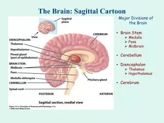
Concept of cone of economy • Normally when humans assume an erect posture, they stand within a certain zone of balance in which their torso remains within a certain distance from their pelvis. • By doing this, energy expended by postural muscles is minimized. • Beyond this zone, the cone of economy, energy expenditure rapidly increases and eventually spinal misalignment results, and external support(cane, crutch, or walker)may be necessary. Dubousset J: Three-dimensional analysis of the scoliotic deformity, in Weinstein SL (ed): The Pediatric Spine: Principles and Practice. New York: Raven Press, 1994, pp 479–496
Concept of cone of economy • Management of spinal deformity includes the recognition and treatment of scoliotic, kyphotic, and spondylolisthetic conditions. • Several radiographic measures have been defined for the assessment of spinal alignment, including coronal, sagittal, and pelvic measures.
Assessment of coronal alignment • The distance between the C-7 plumb line and the central sacral vertical line is defined the amount of coronal plane decompensation in centimeters.
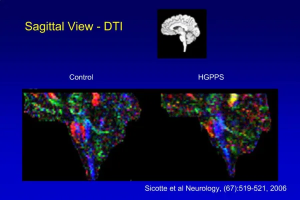
Assessment of sagittal alignment • The most common measure of global SA is the sagittal vertical axis (SVA). • The SVA is measured as the distance between the C-7 plumb line and the posterior superior aspect of the S-1. • SVA: ± 2.5 cm
Spinopelvic inclination • Spinopelvic inclination is a global angular measurement of SA. • It is the angle formed by a line from the femoral heads to the T-1 or T-9 centroid and the vertical plumb line.
Measuringthoracicandlumbarkyphosis • SA is also assessed through regional measures of thoracic and lumbar alignment, including Cobb angles of TK (T2-12 or T5-12), TLK (T10-L2), and LL (T12-S1).
Assessment of pelvicparameters • SVA has traditionally been used to evaluate SA. • Recent data indicate that • The measurement of spinopelvic parameters (PI, PT, and SS) provides a more comprehensive assessment of SA.
Measurements of pelvic incidence • PI is defined as the angle subtended by a line drawn between the midpoint of the sacral endplate to the center of the bicoxofemoral axis and a line drawn perpendicular to the center of the sacral endplate. • Following puberty, PI is generally considered to be a fixed morphological parameter, reflecting the relationship of the sacrum to thepelvis. • PI could be changed (3) by motion of the sacroiliac joint due to degeneration, trauma or iatrogenic injury. Legaye J, et al.: Pelvic incidence: a fundamental pelvic parameter for three-dimensional regulation of spinal sagittal curves. Eur Spine J 7:99–103, 1998 O’Brien MF, (eds): Spinal Deformity Study Group Radiographic Measurement Manual. Memphis, TN: Medtronic Sofamor Danek, 2004
Measurements of pelvic tilt • PT is defined as the angle subtended by a line drawn from the midpoint of the sacral endplate to the center of the bicoxofemoral axis and a vertical plumb line extended from the bicoxofemoral axis. O’Brien MF, Kuklo TR, Blanke KM, Lenke LG (eds): Spinal Deformity Study Group Radiographic Measurement Manual. Memphis, TN: Medtronic Sofamor Danek, 2004
Measurements of sacral slope • SS is defined as the angle subtended by a line drawn along the endplate of the sacrum and a horizontal reference line extended from the posterior/anterior superior corner of S-1 O’Brien MF, Kuklo TR, Blanke KM, Lenke LG (eds): Spinal Deformity Study Group Radiographic Measurement Manual. Memphis, TN: Medtronic Sofamor Danek, 2004
Spinopelvic parameters in asymptomatic subjects • PI: 50.6 (±11.6) • SS: 37.7 (±9.7) • PT: 12.6 (±9.6) • LL (L1-S1): 54.6 (±12.6) • TK: 32.5 (±10.9) • Noshchenko A. et al: Spinopelvic parameters in asymptomatic subjects without spine disease and deformity. Clin Spine Surg 2017 Mar 31. [Epub ahead of print] • Boulay C, et al. Sagittal alignment of spine and pelvis regulated by pelvic incidence: standard values and prediction of lordosis. Eur Spine J 15:415–422, 2006
Pelvic retroversion • As PT increases, the SS decreases because the sacrum assumes a more vertical position about the femoral head axis (pelvic retroversion). • Pelvic retroversion is a compensatory mechanism used to sagittally rebalance the spine and maintain upright posture in cases of SSM.It compensates for decreased LL. • Normal gait requires forward flexion of the pelvis. • Higher pelvic retroversion causes more energy consuming and increasing muscle effort for standing. It is a cause of pain and disability. • Duval K, et al: The mechanical relationship between the rearfoot, pelvis and low-back. Gait Posture 32: 637–640, 2010 • Sarwahi V, et al: Characterization of gait function in patients with postsurgical sagittal (flatback) deformity: a prospective study of 21 patients. Spine (Phila Pa 1976) 27:2328–2337, 2002
Pelvic retroversionfor lumbar stenosis • A portion of the pelvic retroversion can be compensatory for distal lumbar stenosis(L4-S1) as well as for sagittal imbalance. • Lumbar spinal stenosis is significantly associated with the flexible deformities (67%) • Flexible sagittal imbalance is a 10° change in LL between weight-bearing and non–weight-bearing images. Pourtaheri S et al. Pelvic retroversion: a compensatory mechanism for lumbar stenosis. J Neurosurg Spine 27:137–144, 2017
Importance of spinopelvic parameters • A high PI/ PT are a predisposing factor for facet joint degeneration at the lower lumbar spine. • Increased PT values reflected pelvic retroversion and correlated with worsening Health-related quality of life (HRQOL) scores. • T1SPI was more accurately correlated with HRQOL scores than did sagittal vertical axis (SVA). Pourtaheri S et al. Pelvic retroversion: a compensatory mechanism for lumbar stenosis. J Neurosurg Spine 27:137–144, 2017 Lafage V, et al: Pelvic tilt and truncal inclination: two key radiographic parameters in the setting of adults with spinal deformity. Spine (Phila Pa 1976) 34:599–606, 2009
Importance of spinopelvic parameters • The goal of spinal realignment procedures should be an SVA <5 cm, a T1SPI < 0°, and a PT < 20°. • The relationship between PI and LL (spinopelvic harmony) is other important parameter. • LL = PI ± 9°. • Schwab F, et al. Adult spinal deformity-postoperative standing imbalance: how much can you tolerate? An overview of key parameters in assessing alignment and planning corrective surgery. Spine (Phila Pa 1976) 35:2224–2231, 2010 • Lamartin C. Criteria to restore the sagittal balance in deformity and degenerative spondylolisthesis. Eur Spine J 21 (Suppl 1):27–31, 2012
Lack of pelvic compensation for a high SVA • Patients with an elevated SVA and a low PT (lack of pelvic compensation for a high SVA) represent a distinct subgroup in which attempted operative correction is subject to greater risk of postoperative failure. • This pattern may be seen in 1) patients with preexisting hip flexion contracture 2) patients with degenerative flat back with primary extensor muscle pathology 3) globally decompensated patients with secondary extensor muscle weakness 4) patients leaning forward to compensate for severe lumbar stenosis. Ames CP et al Impact of spinopelvic alignment on decision making in deformity surgery in adults . J Neurosurg Spine 16:547–564, 2012
Pelvic obliquity • Pelvic obliquity and associated etiology should be considered in the coronal plane correction strategy. • Pelvic obliquity is quantified by measuring the angle formed between a horizontal reference line and a pelvic coronal reference line.
Pelvic obliquity • Pelvic obliquity due to leg length discrepancy may produce a compensatory lumbar curve to balance the spine. • Correction of this lumbar curve without correction of the primary driver of the pelvic obliquity may lead to coronal decompensation.
Pelvic obliquity • Pelvic obliquity may be secondary(for example, resulting from attempts to compensate for the spinal scoliotic curve). • Curve correction strategies must be sufficient magnitude to allow the pelvis to relax in the coronal plane.
Treatment of patients with scoliosis, coronal misalignment and pelvic obliquity • All patients should be evaluated clinically and radiographically for a leg length discrepancy. • If a leg length discrepancy is identified, the patient should be reevaluated both clinically and radiographically after fitting with a shoe lift to assess how the spine and pelvis respond to correction of the discrepancy.
Treatment of patients with scoliosis, coronal misalignment and pelvic obliquity • Patients with a flexible curve due to pelvic obliquity as a result of a leg length discrepancy may respond well to the addition of a shoe lift only or surgical treatment of the leg length discrepancy.
Summary • The pelvis plays a critical role in balanced upright sitting and standing postures. • Treating physicians must evaluate pelvic alignment, including PI, PT, SS, and pelvic obliquity, as well as more traditional measures such as SVA, LL, TK, and regional scoliotic curves. • Particular attention must be paid to PT, a dynamic pelvic parameter reflecting pelvic retroversion, since increased PT implies residual postoperative spinal deformity and negatively affects function and thus postoperative outcomes.
Treatment of patients with scoliosis, coronal misalignment and pelvic obliquity • If the spinalcurve is rigid, it will not be corrected after the addition of a shoe lift. • In this case, there are 2 options: • 1) to correct the curve perpendicular to the oblique pelvis and ignore the pelvic obliquity, or • 2) to correct the spine to a level pelvis if leg length correction is planned (for example, with a future hip replacement) or if the patient tolerates a shoe lift.
Preoperativeradiologicalevaluation • Comparison of preoperative standing and prone radiographs may assist the surgeon in estimating the anticipated difference in alignment likely to be seen on the operative table and therefore the likely magnitude of optimal curve correction. • Degenerative deformity may reduce up to 30% in the supine position. • Preoperative bending radiographs will also assist the surgeon in determining how much correction may occur if a curve is not included in the fusion.
Location of the osteotomy • The location of the osteotomy along the spine is also an important consideration when attempting to normalize PT. • More caudal osteotomies in the lumbar spine are significantly correlated with greater PT reductions, and the degree of osteotomy is significantly correlated with changes in TK. • Lafage V, et al: Multicenter validation of a formula predicting postoperative spinopelvic alignment. Clinical article. J Neurosurg Spine 16:15–21, 2012 • Lafage V, et al: Spino-pelvic parameters following surgery can be predicted: a preliminary formula and validation of standing alignment. Spine (Phila Pa 1976) 36:1037–1045, 2011 • Smith JS, et al: Dynamic changes of the pelvis and spine are key to predicting postoperative sagittal alignment following pedicle subtraction osteotomy. Spine (Phila Pa 1976).37(10):845-53, 2012
Assessment of location for PSO • The angle subtended between the patient’s spine and the planned C-7 plumb line (which are thinner white lines) represents the angle of correction needed given that the PSO is at that level. • Thus, as the PSO moves more caudal, the angles become smaller. • Blue line represents the C-7 plumb line • White vertical line shows approximate corrected location of C-7 plumb line • Thick angled white line, the patient’s spine.
Calculation of the amount of correction needed • Lamartina et al. proposed the spinofemoral angle (SFA) to calculate the amount of correction needed in sagittal imbalance surgery. • This method is based on a single angle, formed by the femoral axis and the line drawn from the center of C7 to the point where the vertical line from the posterior end of S1 plate intersects the level of planned osteotomy. • To increase the accuracy of this method, it is possible to add two parameters: hip extension reserve and thoracic hypokyphosis. • Needed correction angle = SFA + 10 +increase in thoracic kyphosis (active flexion TK - TK) Lamartina C, et al. Criteria to restore the sagittal balance in deformity and degenerative spondylolisthesis. Eur Spine J 21(Suppl 1):S27–S31, 2012
Sagittalandcoronalbalance • Historically, Treatment has focused on scoliosis correction and the prevention of scoliotic curve progression. • However, Recent data have demonstrated the impact that sagittal plane deformities and global spinal alignment have in the generation of pain and disability. • Consequently, Increased emphasis has been placed on restoring physiological LL, TK, and the C-7 plumb line (sagittal vertical axis-SVA). • Treating physicians must be familiar with the radiographic findings consistent with sagittal spinal misalignment (SSM).
PI: 55.48 (±5.31) • SS: 35.99 (±7.53) • PT: 17.97 (±7.16) • LL: 48.84 (±9.82) • TK: 32.55 (±10.92) Sudhir G et al. Radiographic Analysis of the Sacropelvic Parameters of the Spine and Their Correlation in Normal Asymptomatic Subjects. Global Spine Journal Vol. 6 No. 2/2016
General algorithm for the treatment of patients with scoliosis, coronal misalignment and pelvic obliquity • In patients with pelvic obliquity in which the spine is flexible and aligned, full correction of the curve may lead to significant coronal decompensation, as the scoliotic curve may be compensatory. • In some of these patients, the addition of a shoe lift will allow the flexible spine to relax and may improve alignment and deformity-related symptoms. • If the spine is rigid, as is more common in adults, a shoe lift may be poorly tolerated and may not be effective in rebalancing the spine. • Patients in such cases may require incomplete curve corrections and sometimes a shoe lift as well depending on the final standing alignment. • PO = pelvic obliquity.
Drawings of complex curve patterns • Drawings of complex curve patterns in which • the coronal correction strategy must take into account the patients probable alignment shift when standing to determine how much curve correction is possible in each direction. • Often this may involve shifting the patient’s coronal plumb slightly off to the side opposite the short leg.
spinopelvic balance • Spinopelvic balance is determined by the association of pelvic alignment with the lumbar spine. • In this geometric construction, the superior angle of the S1 endplate with a horizontal line (sacral slope) is equal to the lower lumbar lordosis angle. • Spinopelvic balance is measured as the difference between the excess of lordosis measured in a patient and the predicted excess of lordosis, calculated from pelvic incidence and age-related spinopelvic constant. • spinopelvic balance is between pelvic incidence and difference of thoracic kyphosis and lumbar lordosis, excess of pelvic tilt, knee flexion and thoracic compensatory hypokyphosis. • spinopelvic balance in the interval (0, +10) is better outcomes • Best scores is found in patients with both spinopelvic balance (0, +10) and sagittal balance (measured as deviation of C7 plumbline from S1).