Download
1 / 41
410 likes | 483 Views
The Blood and Endocrine System. By: Ashley Hahn Daniel Ron Dylan Angeles Adrian Garcia Period 3. Hematopoiesis. The production of all types of blood cells Originate as stem cells Some of these daughter cells stay as these stem cells in order to prevent depletion.

E N D
The Blood and Endocrine System By: Ashley Hahn Daniel Ron Dylan Angeles Adrian Garcia Period 3
Hematopoiesis • The production of all types of blood cells • Originate as stem cells • Some of these daughter cells stay as these stem cells in order to prevent depletion. • Cells become either myeloid (all cells except WBCs) and lymphoid progenitor (WBC) cells. • Amount of each is regulated by the bone marrow • 3 paths: Erythroid cells, lymphocytes, myelocytes • Blood counts are important because they determine the amount of blood and how much of the blood components are in the blood to help diagnose diseases.
Erythropoiesis • Process in which red blood cells are produced. • Stimulated by lack of oxygen in circulation, which causes the kidneys to secrete the erythropoietin, which causes more stem cells to be differentiated to cells that will mature to red blood cells.
This is the process of differentiation in erythropoiesis: • Hemocytoblast a multipotent hematopoietic stem cell • Common myeloid progenitor • Unipotent stem cell • Pronormoblast also commonly called proerythroblast or rubriblast. • Basophilic normoblast/early normoblast also commonly called erythroblast • Polychromatophilic normoblast/intermediate normoblast • Orthochromatic normoblast/late normoblast - Nucleus is expelled before becoming a reticulocyte (released from the bone) • Reticulocyte (step from red blood cell) • In about 2-3 days the reticulocyte turns into a mature red blood cell. • Stay in circulation for about 100-120 days, then attacked by macrophages.
Erythrocytes (RBCs) • Main Function: Transport oxygen from the lungs to living tissue and carry away carbon dioxideas well as regulating pH levels. • Regulations of core body temperature (ex. vasodilation/contraction) • Produced in red bone marrow from stem cells • Contains NO nuclei • 40-50% of total blood volume • Contains hemoglobin (makes up 95% of RBCs), which is a gas transporting protein molecule.
Iron in Hemoglobin • Iron(II) tends to exist in a high spin configuration where unpaired electrons exist in Eg antibonding orbitals. • Iron (III) has an odd number and must have one or more unpaired electrons to become more stable. • Fe 2+ binds to a single oxygen, Fe 3+ binds to O2- and Fe 4+ binds to peroxide O2^2- • Due to its high reactivity and the oxygen in the blood, there is a lot of iron in your blood.
Leukocytes (WBCs) • Main Function: • Seek out, identify, and bind to alien protein on bacteria, viruses, and fungi so that they can be removed by granulocytes and macrophages (also gets rid of dead or dying cells and foreign matter). • Only about 1% of total blood volume (in healthy people) • Most produced in bone marrow from stem cells, others in the liver, spleen, thymus and lymph glands. • A response to foreign particles invading the body.
Thrombocytes (platelets) • Main function: • Works with blood clotting chemicals • Cell fragments without nuclei • Can release coagulating chemicals which cause clots to form that can plug up narrowed blood vessels. • Aid in fighting infection by releasing proteins that kill invading bacteria • 1/3 the size of a RBC • Also produced in bone marrow from stem cells
Plasma • Main function: • Transports nutrients to the cells and carries RBCs, WBCs, and platelets throughout the body. • Clear, yellow tinted water (92+%), sugar, fat, protein, and salt solution. • 55% of total blood volume • Contains blood clotting factors, sugars, lipids, vitamins, minerals, hormones, enzymes, antibodies, and other proteins. • Approximately 500 body produced proteins have been identified in plasma.
Coagulation • An important part of hemostasis: • Hemostasis is the process of repairing a damaged blood vessel. • Disorders can lead to increased bleeding, called hemorrhage, or obstructive clotting, called thrombosis. • Begins immediately after injury • Four major steps: • Vasoconstriction • Platelet plug • Blood clot formation • Dissolving clot
Coagulation Cont. - Endothelium • Coagulation begins when the endothelium lining is damaged. • A thin layer of cells that lines the interior surface of blood vessels and lymphatic vessels. • Functions include: • Semi-selective barrier • Blood clotting (thrombosis and fibrinolysis) • Inflammation • Angiogenesis • Vasoconstriction and vasodilation (control of blood pressure)
Extrinsic Pathway • First pathway activated • Begins from disrupted vessel wall • Damage to tissue; protein: tissue factor (TF) • Tissue factor (factor III) activated by: • Cytokines • Cell injury • Vessel disruption (blood in tissue) • External cells come into contact with blood factor VII activated into VIIa leads to factor X • Factor X Prothrombin Thrombin Fibrinogen Fibrin
Intrinsic Pathway • Begins with platelet plug • Endothelial cells produce Von Willebrand factor (vWF), a glycoprotein • When endothelial layer is injured, collagen, vWF, and TF are exposed to the bloodstream. • Platelet contact with collagen or vWF = activated platelets • Intrinsic pathway begins in factor XII (Hageman factor) and leads to factor X. • Factor X Prothrombin Thrombin Fibrinogen Fibrin • Platelet aggregation uses fibrin to clump • Forms clot
Fibrin and Thrombin • Fibrin forms the meshwork of a blood clot. • It’s laid over the platelet plug and traps blood cells and other particles in to secure the clot. • Fibrinogen (Factor I) • Inactive form of fibrin in the bloodstream • Requires thrombin to be activated • Prothrombin (Factor II) • Inactive form of thrombin in the bloodstream • Required prothrombin activator to be converted into thrombin • Prothrombin Thrombin Fibrinogen Fibrin Platelets clot formed
Limiting the Clot Size • Coagulation Inhibitors (stops growth): • Interferes with fibrin formation • Done by antithrombin III, protein S, and protein C. • Fibrinolysis (breaks down clot): • Plasminogen activators are slowly released from the inner lining of the damaged vessel wall; dissolves part or the entire clot. • When completely healed, the rest of the clot is dissolved.
Coagulation Summary • Process of coagulation: • Damaged vessel wall • Vessel constricts to reduce flow of blood • Platelet adhesion • Activated platelets release chemicals to keep vessel constriction and attract more platelets • Platelet aggregation • Tissue factor sets in motion of the clotting factors floating in the blood • Meshwork formed • RBCs and WBCs get trapped in the plugs • Coagulation inhibitors or fibrinolysis takes place • Damaged wall heals and clot is dissolved
Coagulation Causes • Causes of blood clots include: • Inactivity for long periods of time • Smoking Recently having surgery or an injury • Obesity • Heart defects • Atrial fibrillation: uncoordinated quivering causes the heart to pump blood out inefficiently • Damaged valves • Walking for long periods of time (blood pools in the legs)
Prevention Considerations • Physical activity • Sitting down after long walks • When traveling for long periods of time, stop for breaks or walk when possible. • Sitting: bend the toes toward the knees and relaxing them several times or lifting the heels and pressing down on the toes to contract leg muscles. • Heparin, an anticoagulant, is a treatment for people with blood clots.
Blood Typing • Depends on whether or not there are certain proteins (antigens) on your RBCs. • Four categories: • Type A • Receives types A and O blood • Type B • Receives type B and O blood • Type AB • Receives type A, B, AB, and O blood • Type O (universal blood donor) • Receives only type O blood
ABO and Back Typing • ABO Typing: If your blood cells stick together (agglutinate) when mixed with: • Anti-A serum, you have type A blood • Anti-B serum, you have type B blood • Both anti-A and anti-B serum, you have type AB blood • Neither anti-A or anti-B serum, you have type O blood • Back Typing: If your blood clumps together when: • B cells are added to your sample, you have type A blood • A cells are added to your sample, you have type B blood • Either of cells are added to your sample, you have type O blood *Lack of cells sticking together indicates you have type AB blood
RH Typing • If your blood cells: • Stick together when mixed with anti-Rh serum, you are Rh-positive. • Receives only Rh-positive blood • Do not stick together when mixed with anti-Rh serum, you are Rh-negative. • Receives only Rh-negative blood
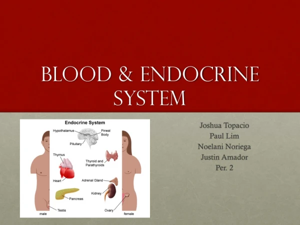
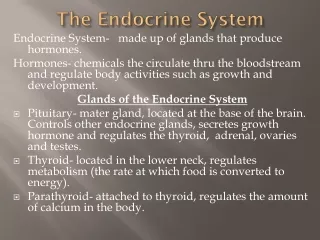
Endocrine System • A system of glands which secret hormones into the internal environment (body fluids). • Hormones spread from the body fluids into the bloodstream and later act on target cells.
Glands • Glands help to regulate the metabolic processes: • Chemical reactions • Water balance • Substances • Electrolytes • Blood pressure • They also play a part in reproduction, development, growth, and homeostasis.
Glands Cont. • Hypothalamus: Located below the thalamus, above the brain stem. • CRH • GnRH • Somatostatin • GHRH • DA • TRH
Glands Cont. • Pituitary Gland “master gland”: Located in the sella turcica. • Anterior Pituitary Gland • FSH • GH • LH • PRL • TSH • Posterior Pituitary Gland: • ADH • Oxytocin
Glands Cont. • Thyroid Gland: Located in the neck, below the thyroid cartilage. • Calcitonin • T4 • T3 • Parathyroid Gland: Located in the neck. • Parathyroid hormone
Glands Cont. • Adrenal Medulla: Located at the center of the adrenal gland. • Epinephrine • Norepinephrine • Adrenal Cortex: Located along the perimeter of the adrenal gland. • Aldosterone • Cortisol
Glands Cont. • Pancreas: Lies posterior to the stomach and is attached to the small intestine. • Glucagon • Insulin • Somatostatin • Pancreatic Polypeptide
Glands Cont. • Pineal Gland: Attached to the thalamus near the roof of the third ventricle. • Melatonin • Thymus Gland: Lies posterior to the sternum and between the lungs. • Thymosin
Glands Cont. • Liver: Lies below the diaphragm. • Somatomedin • Angiotensinogen & Angiotensin • THPO • Hepcidin
Control Mechanisms • As hormone levels rise in the blood and the hormone exerts its effects, negative feedback inhibits the system because of the body's need for homeostasis. • It inhibits the system and hormone secretion decreases. • As hormone levels in the blood decrease and the hormone effects decline, inhibition of the system stops and secretion of the hormone increases again.
Hormone Levels Too High or Low • Process of high level: • Hormone levels rise • Hormone control mechanism senses change • Endocrine gland is inhibited • Hormone secretion is decreased • Hormone levels return to normal • Process of low level: • Hormone levels drop • Hormone control mechanism senses change • Endocrine gland is stimulated • Hormone secretion is increased • Hormone levels return to normal
Bibliography Barnes, Broda. “Endocrine System.” Research Foundation Inc. 10 Dec. 2012 <http://www.brodabarnes.org/endocrine_sys.htm.> “Blood Typing.” Medline Plus. National Institutes of Health. 27 Feb. 2013 <http://www.nlm.nih.gov/medlineplus/ency/article/003345.htm> “Definition of Hematopoiesis.” MedicineNet Inc. 2 Mar. 2013 <http://www.medterms.com/script/main/art.asp?articlekey=19775> Doohan, James. “Hemostasis.” Biological Sciences. Human Physiology. 11 Oct. 1999 <http://www.biosbcc.net/doohan/sample/htm/Hemostasis.htm> "Endocrine System." Wikipedia. Wikimedia Foundation, 16 Feb. 2013. Web. 16 Feb. 2013. <http://en.wikipedia.org/wiki/Endocrine_system>. O’Neil, Dennis. “Blood Components.” Behavioral Sciences Department. <http://anthro.palomar.edu/blood/blood_components.htm> Shier, David, Jackie Butler, and Ricki Lewis. Hole''s Human Anatomy & Physiology. 11th ed. New York: McGraw-Hill, 2007. Print.