Download
1 / 50
610 likes | 1.08k Views
1. Stomach and 2. esophageal cancer. Stomach cancer-Anatomy. Parts of the stomach: -cardia (cardiac portion) -fundus -body -pyloric antrum -pyloric canal. -cancer on the lesser curvature is more frequent than on the greater

E N D
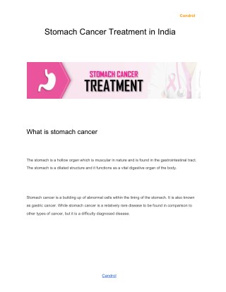
Stomach cancer-Anatomy • Parts of the stomach: -cardia (cardiac portion) -fundus -body -pyloric antrum -pyloric canal -cancer on the lesser curvature is more frequent than on the greater - cancer in the body or antrum more frequent but cancer in the cardiac region is increasing rapidly the Western world (still, everywhere, body and antrum are more frequent)
Epidemiology • Adenocarcinoma of the stomach was the leading cause of cancer-death worldwide through most of the 20th century (1901-2000) -now ranks second to lung cancer • It’s incidence had an ever more marked decline in North-America and Western-Europe
In Romania: -second cause of cancer-death in males -fifth in females
Etiology (risk factors) • Genetic factors Hereditary diffuse gastric cancer (E-cadherin mutations, autosomal dominant disease)Blood type A Pernicious anemia Familial adenomatous polyposis (FAP), Hereditary nonpolyposis colon cancer Li-Fraumeni syndrome BRCA1/2 mutations Family history (other, yet unidentified genetic factors) II. Precursor lesions Adenomatous gastric polyps Chronic atrophic gastritis Dysplasia Intestinal metaplasia Menetrier's disease
Etiology (risk factors) III. Environmental factors Helicobacter pylori infection Nutritional High salt consumption High nitrate consumption Low dietary vitamin A and C Poor food preparation (smoked, salt cured) Lack of refrigeration (mycotoxins) Poor drinking water (well water: nitrates)Occupational Rubber workers Coal workersCigarette smokingEpstein-Barr virusRadiation exposurePrior gastric surgery for benign gastric ulcer disease [scaring and regurgitation of the (irritant) bile]
Histological subtypes-Lauren’s classification • Adenocarcinoma • Intestinal type -microscopically: gland formation -related with H. pylori -related to precancerous conditions: chronic gastritis, atrophy, intestinal metaplasia -increasing incidence with age -men>women • Diffuse type -less well differentiated, characterized by sheets of cells without gland formation, with the occasional presence of signet ring cells and mucin -related with H. pylori, but genetic factors more important -not related to the above precancerous conditions -mostly younger patients -women>men -worse prognosis
Intestinal type Chronic gastritis with intestinal metaplasia: The mucinous gastric epithelium (arrowhead) is replaced by intestinal type of epithelium (arrow). Gastric cancer-intestinal type
Macroscopic subtypes-Bormann’s classification • Bormann type 1: polypoid or protuberant carcinoma (usually a well-differentiated adenocarcinoma) • Bormann type 2: expansive ulcerating carcinoma (deep ulcer with elevated margins) • Bormann type 3: infiltrative ulcerating carcinoma • Bormann type 4: diffusely infiltrating carcinoma= linitis plastica. • Bormann type 5-unclassifiables • The macroscopic appearance is of prognostic value: higher the Borrmann type number, worse the prognosis
Extension of stomach cancer-direct extension • Greater omentum • Lesser omentum =hepatogastric ligament • Colon • Pancreas • Duodenum • Cardiac tumors (stomach extraperitoneal): diaphragm
Extension of stomach cancer-peritoneal extension • possible after a lesion extends beyond the gastric wall to a free peritoneal (serosal) surface • Krukenberg tumor (mucin-secreting signet-ring cell metastasis on the ovary from gastrointestinal or breast cancers) • Blumer’s shelf (i.e. shelf-like tumor of the anterior rectal wall)
Extension of stomach cancer-lymph vessels->lymph nodes • Regional lymph nodes: mainly along the arteries: -lesser and greater curvature -celiac -splenic hilum -hepatic hilum -pancreatico-duodenal -some paraaortic (in the Japanese staging) • Lymph nodes where spread is considered metastasis: -paraaortic -mediastinal -left supraclavicular (Virchow's) -left axillary (Irish) -umbilical (Sister Mary Joseph's)
Extension of stomach cancer-hematogenic metastases • Liver • Lung • Bone • Brain
Clinical features • Late (cancer limited to the stomach in about 15% of patients) • Nonspecific signs and symptoms: -Weight loss -Anorexia (sometimes selective anorexia to meat) -Abdominal pain (can be similar to that in ulcer) -Anemia secondary to chronic blood loss
Clinical features Proximal cancer: -dysphagia Distal cancer: - gastric outlet obstruction: nausea, vomiting Linitis plastica: -early satiety (decreased gastric capacity) Other signs and symptoms: -hematemesis (rarely) -left supraclavicular adenopathy, left axillary adenopathy -paraneoplastic syndromes: venous thrombosis etc.
Diagnosis • Barium Meal -Better tolerated -Sensibility: only 50% • Upper GI endoscopy-gold standard -Sensitive and specific -can perform biopsy -more expensive
Diagnosis • CT or better, MRI of the abdomen (lymph nodes and peritoneal/hepatic metastases) • EUS (endoscopic ultrasonography) can help decide resectability • Pulmonary radiography • High resource setting: PET/CT
Staging Gastric cancer is a surgically staged disease -TNM staging Tis-in situ tumor T1-invading the lamina propria or the submucosa T2-muscularis propria/subserosa T3-serosa T4-adiacent organ involvement N0: no positive lymph nodes N1: 1-6 lymph nodes positive N2: 7-15 N3: more than 15
Treatment RESECTABLE TUMORS • Tis and T1 tumors limited to de mucosa (T1a) can be managed by endoscopic mucosal resection • T1b-T3 N+/- : gastric resection with at least 4 cm margin (- Wedge resection -Segmental resection -Proximal gastrectomy -Pylorus preserving gastrectomy -Distal gastrectomy -Total gastrectomy) • T4 tumors require en bloc resection of invaded structures • Gastric resection should include D1+D2 lymph node resection (D1=perigastric lymph nodes; D2=those along the named arteries of celiac axis. At least 15 lymph nodes have to be excised.) HE staining for lymph nodes is not enough; misses 50% of lymph node meta=>additional cytocheratin staining is used
Treatment RESECTABLE TUMORS After surgery in T3-T4 N0-N1 patients: In Europe and America: -usually there is no correct D2 resection STANDARD: adjuvant chemo-radiotherapy In Japan: -usually there is a correct D2 resection STANDARD: adjuvant oral chemotherapy plus immunostimulating Coriolus versicolor extract -one time intraperitoneal chemotherapy after surgery might be used (with cisplatin)
Treatment UNRESECTABLE TUMORS -chemoradiotherapy reevaluation and if operable=> surgery Not operable after chemoradiotherapy: -obstructive symptoms: palliative gastric resection or gastro-jejunostomy -chemotherapy
Helicobacter pylori eradication-mandatory after primary treatment (for example subtotal gastric resection) • First line: triple therapy: -Clarithromycin 3x 500 mg/day -Amoxicillin 2x1000 mg/day -Omeprazole 1x20 mg/day • Second line: metronidazole based regimen • Third line: e. g. Furazolidon
Side effects of gastric surgery • Total gastrectomy=>loss of intrinsic factor which binds vitamin B12=>IM B12 supplementation/PO B12+intrinsic factor • Some patients can not tolerate complete gastrectomy: if nutrition is inadequate they will die of malnutrition • Dumping syndrome: early and late
Under normal physiologic conditions the stomach controls the rate at which the gastric contents leave the stomach. I. Early dumping syndrome: due to quickly filling of the jejunum with undigested food from the stomach • =>bowel distension • => movement of water from the blood to the intestine to dilute the intestinal contents Symptoms: abdominal bloating, pain/cramping, vomiting, flushing, sweating, rapid heart rate, light headedness and diarrhea. II. Late dumping syndrome: hypoglycemia due to increased insulin secretion as a response to high serum glucose peak The small bowel is very effective in absorbing sugar, so that the rapid absorption of a relatively small amount of sugar can cause the glucose level in the blood to rise rapidly. The pancreas responds to this glucose challenge by increasing the insulin output. Unfortunately, the sugar that started the whole cycle was such a small amount that it does not sustain the increase in blood glucose, which tends to fall back down at about the time the insulin rush starts. Symptoms: fatigue, sleepiness
Screening of gastric cancer • In the Western world screening with endoscopy is low yield • But even here there are high risk groups that may benefit from stomach cancer screening: • Older people with gastritis or pernicious anemia • Partial gastrectomy • Polyps in the stomach • Genetic predisposing conditions [Familial adenomatous polyposis (FAP), Hereditary nonpolyposis colon cancer (HNPCC)] • Immigrants from countries where stomach cancer is more common.
Screening of gastric cancer • In Asia and other high incidence countries: screening with endoscopy would be a reasonable choice if economically sustainable • Screening programs: Japan, South-Korea Case-control studies suggested a 40–60% decrease in gastric cancer mortality with photofluorography (barium meal) screening in Japan and Venezuela
Questions • What are the risk factors for gastric cancer? • What are the two main histological types of gastric cancer and enumerate some differences. • What are the symptoms of gastric cancer? • What diagnostic tools should be used for diagnosis of gastric cancer? • What is the main treatment type for gastric cancer and how it is done? • What is the early and late dumping syndrome?
Anatomy of the esophagus • From the inferior margin of the cricopharyngeus muscle (or from the inferior margin of the cricoid cartilage) • To the cardia of the stomach • ~25 cm in length
Subdivision of the esophagus There are two subdivision systems for the esophagus and they are not equivalent Subdivision system 1 • Cervical-begins at the lower end of pharynx (level of 6th vertebra or lower border of cricoid cartilage) and extends to the thoracic inlet (suprasternal notch); 18 cm from incisors. • Thoracic -Upper thoracic: from thoracic inlet to level of tracheal bifurcation; 18-23 cm. -Mid thoracic: from tracheal bifuraction midway to gastroesophageal junction; 24-32 cm. -Lower thoracic: from midway between tracheal bifurcation and gastroesophageal junction, including abdominal esophagus; 32-40 cm. • Abdominal-Considered part of lower thoracic esophagus; 32-40 cm. Subdivision system 2 • Upper third (10% of esophageal cancers) • Middle third (40%) • Lower third (50%)
Normal histology • It is lined with stratified non-keratinizing squamous epithelium • The lower third (5 to 10 cm) of the esophagus may contain glandular elements. Replacement of the stratified squamous epithelium with columnar epithelium is referred to as Barrett's esophagus
Normal histology • The lower third (5 to 10 cm) of the esophagus may contain glandular elements. Replacement of the stratified squamous epithelium with columnar epithelium is referred to as Barrett's esophagus
Histological subtypes of esophageal cancer • 90% squamous cell carcinoma • 10% adenocarcinoma
Epidemiology • Rare cancer in North America and Europe • High frequency in northern China, Iran, India and near the Caspian Sea [alkaline soil, ingestion of nitrosamines and low riboflavin (=vitamin B2), nicotinic acid, Mg and Zn]
Risk factors for squamous cell carcinoma • In North America and Western Europe, alcohol and tobacco use are the major risk factors for squamous cell carcinoma, accounting for 90% of cases • Other risk factors: -HPV infection -nitrate-rich foods (pickled vegetables, alcoholic beverages, cured meats and fish) -Plummer-Vinson (Paterson-Kelly) syndrome= iron deficiency anemia + low riboflavin; increased risk for esophageal and oral cavity + hypopharyngeal cancers
-achalasia (failure of the lower esophageal sphincter to relax during swallowing) -caustic burns (especially lye corrosion) -tylosis (hyperkeratosis of the palms and soles and papilloma of the esophagus)
Risk factors for adenocarcinoma • Barrett's esophagus (caused by severe and long-standing gastroesophageal reflux disease (GERD) • Obesity and hiatal hernia-possibly due to an increased risk of reflux • Smoking
Extension • DIRECT EXTENSION: No serosa is present, facilitating extra-esophageal spread of disease. (The four esophageal layers: an innermost epithelial layer, inner circular muscle layer, an outer longitudinal muscle layer and an adventitia.) trachea, bronchia, pleura, lung, pericardium, large vessels, recurrent nerves, diaphragm
Extension • Lymphatic spread -cervical esophagus->upper mediastinal, inferior cervical, supraclavicular -thoracic esophagus->mediastinal -lower thoracic=abdominal esophagus-> mediastinal, celiac • Metastases: -liver -lung -suprarenals -bone
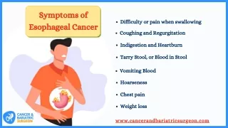
Clinical features • Dysphagia=difficulty in swallowing -first for solids, then for liquids • Odynophagia=painful swallowing • Invasion/compression of the trachea: cough, dyspnea, hemoptisis • Invasion of the recurrent nerve/nerves: dysphonia • Nonspecific signs and symptoms: -Weight loss -Anorexia
Diagnosis and evaluation of extension • Upper digestive endoscopy with biopsy • Endoscopic US (for evaluation of local extension and lymph node metastases) • Barium swallow (no indication if endoscopy is done first) • Head and neck exam (for lymph node metastases) • CT or better, MRI, or even better PET/CT of the thorax, cervical region, upper abdomen • General evaluations for an eventual surgery
Treatment • Tis and T1a (tumor invades lamina propria) =>endoscopic mucosal resection • T1bN0 (tumor invades submucosa) =>esophagectomy • All other loco-regional disease (T1bN1, T2-T4, N0-N1, M0-M1a): A) For squamous cell carcinoma: Chemoradiation -> reevaluation* ≥5 weeks: persistent disease=>salvage surgery or boost chemoradiation *Reevaluation by CT with contrast, PET/CT, endoscopic US, esophageal endoscopy
Treatment B) For adenocarcinoma: Chemoradiation -> esophagectomy + lymphadenectomy
Questions • What are the two main types of esophageal cancer and what risk factors do they have? • What are the symptoms of esophageal cancer? • What is the treatment for locally advanced esophageal cancer? (Treatment for both squamous cell carcinoma and adenocarcinoma should be described.)