Download
1 / 26
260 likes | 271 Views
5 th presentation. Radiographic technique of Pelvis, hip joint and sacroiliac joint. Pelvis, hip joint and sacroiliac joint. AP Pelvis (Bilateral hips) Basic. Film Size : HD 35x43 cm14x17in (crosswise).

E N D
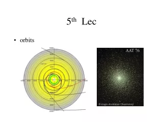
5th presentation Radiographic technique of Pelvis, hip joint and sacroiliac joint
AP Pelvis (Bilateral hips) Basic • Film Size : HD 35x43 cm14x17in (crosswise). • *SHIELDING: Shield gonads carefully without obscuring essential anatomy. • Patient Position: Patient supine. • Part Position : arms at sides or across chest, pillow under head, support under knees, separate legs and, feet and then internally rotate feet and lower limbs 15 to 20,sandbag between heels, feet taped together . Distance: 100 cm or 40 in) . CR : perpendicular to the film. C P: Midway between level of ASISs and symphysis pubis 2in (5 cm) superior to the symphysis pubis). NB/ (suspend respiration during Exposure ). Gonadal shielding for males or females without obscuring the essential anatomical parts. Collimation: Collimate on four sides to area of interest.
Structure shown: pelvic girdle, L5, sacrum and coccyx, femoral heads, neck and greater trochanters are visible .
Basic/ AP pelvis (bilateral hips) 1. Lateral part of the sacrum2. Gas in colon3. Ilium4. Sacroiliac joint5. Ischial spine6. Superior ramus of pubis7. Inferior ramus of pubis8. Ischial tuberosity9. Obturator foramen 10.Intertrochanteric crest 11. Pubic symphysis 12. Pubic tubercle13. Lesser Trochanter14. Neck of femur15. Greater Trochanter16. Head of femur17. Acetabular fossa18. Anterior inferior iliac spine19. Anterior superior iliac spine20. Posterior inferior iliac spine21. Posterior superior iliac spine22. Iliac crest
AP Pelvis (Bilateral frog-leg position) – (non-trauma hips) B Film Size: HD 35x43 cm 14x17in (crosswise). *SHIELDING: Shield gonads carefully without obscuring essential anatomy. *Patient Position: Patient supine. Part Position: arms at sides or across chest, both knees flexed 90, femurs abducted 40to 45 from vertical, feet soles placed together ,ensure pelvis is not rotated . Distance:(100 cm or 40 in). C P: 3 in (7.5cm )below level of ASIS (1 in or2.5cm above the symphysis pubic.) CR : perpendicular to the film. Collimation: Collimate on four sides to area of interest. NB / (suspend respiration during Exposure ).
Structure shown: femoral heads, neck and trochanters acetabulum are visible
AP Axial Pelvis (Pelvic outlet) S • Film Size : HD 24 x 30 cm,10x12in crosswise. • *SHIELDING: Shield gonads carefully without obscuring. essential anatomy. • Patient Position: Patient supine. • Part Position: arms at sides or across the chest, pillow for the head, legs fully extended. • Distance: 100 cm or 40 in). • CP: 1-2 in(3-5 cm)distal to superior border of symphysis pubis, or greater trochanters. • CR: 20 - 35 for (males) or 30- 45 for (females) cephalic. • Collimation: Collimate on four sides to area of interest. • NB/ (suspend respiration during Exposure ).
superior ramus of pubes Structure shown: superior and inferior rami of pubes and body and ramus of ischium are visible with minimal foreshorting.
R&L PO Pelvis (for acetabulum) Judet method S Film Size: HD24 x 30 cm, 10x12in crosswise. *SHIELDING: Shield gonads carefully without obscuring essential anatomy. *Patient Position: Patient in semi-supine. Part Position: pillow for head, affected side up or down depending on anatomy to be demonstrated, place patient in a 45 posterior oblique ,support with wedge sponge. femoral head and the acetabulum of interest in the midline of couch. Distance: 100 cm or 40 in. CR: perpendicular to the film. C P: when anatomy of interest is downside, centered to 2 in(5cm) distal and 2in (5cm) medial to downside ASIS. when anatomy of interest is upside, centered to 2 in directly distal to upside ASIS . Collimation: Collimate on four sides to area of interest.
Structure shown: when center to the downside acetabulum, the anterior rim of acetabulum. And the posterior iloischial column are demonstrated. the iliac wingis also well visualized When center to the upside acetabulum, the posterior rim of acetabulum. And the anterior iloischial column are demonstrated . The Obturator foramen is also visualized.
AP Unilateral Hip (+ proximal femur)B Film Size : HD24 x 30 cm, 10x12in lengthwise. SHIELDING: Shield gonads carefully without obscuring essential anatomy. Patient Position: Patient supine. Part Position: arms by the side or over chest, pelvis not rotated,the affected leg rotated medially 15 to 20. Distance:100 cm or 40 in. CP : 1 or 2 in (2.5cm to 5cm) distal to mid femoral neck. CR: perpendicular to the film. Collimation: Collimate on four sides to area of interest.
Structure shown: the proximal one third of the femur should be visualized, along with the acetabulum and adjacent parts of the pubis,ischium, andIlium
Radiographic Anatomy - AP Hip 1 - Anterior superior iliac spine 2 - Ilium 3 - Anterior inferior iliac spine 4 - Pelvic brim 5 - Acetabular fossa 6 - Head of femur 7 - Fovea 8 - superior ramus of pubis 9 - Obturator foramen 10 - Inferior ramus of pubis 11 - Pubic symphysis 12 - Ischium 13 - Lesser trochanter 14 - Intertrochanteric crest 15 - Greater trochanter 16 - Neck of femur
Axiolateral inferosuperior hip (trauma hip) B Film Size : HD 24 x 30 cm, 10x12in lengthwise. SHIELDING: Shield gonads carefully without obscuring essential anatomy. Patient Position: Patient supine. Part Position: unaffected leg flexed and elevated such that thigh is near vertical, and outside the radiation field, cassette placed in the crease above iliac crest 90 to CR, cassette vertical, affected leg rotated 15 - 20 medially unless contraindicated by possible fracture . Distance: 100 cm or 40 in. C P: to the femoral neck . CR: perpendicular to the film. Collimation: Collimate on four sides to area of interest.
Structure shown: entire femoral heads, and neck trochanters ,acetabulum should be visualized.
Unilateral frog-leg hip(non-trauma hip) To show lateral hip joint B Film Size : HD 24 x 30 cm, 10x12in crosswise. SHIELDING: Shield gonads carefully without obscuring essential anatomy. Patient Position: Patient supine. Part Position: hip and knee of affected side flexed, sole of foot against inside of the opposite leg, (near the knee), femur abducted 45 from the vertical. Distance: 100 cm or 40 in. C P: Mid-femoral neck. CR: perpendicular to the film. Collimation: Collimate on four sides to area of interest.
Structure shown: lateral view of acetabulum, and femoral heads, and neck) trochanters area, and proximal one third of the femur should be visualized
AP Axial Sacroiliac joints B Film Size: HD 24 x 30 cm,10x12in lengthwise. SHIELDING: Shield gonads carefully without obscuring essential anatomy. Patient Position: Patient supine. Part Position: pillow for head, legs fully extended, a support under knees. Distance: 100 cm or 40 in C P: 2 in (5cm) below the level of ASIS. CR: 30- 45 cephalic (30 for males, 35 for females). Collimation: Collimate on four sides to area of interest.
Structure shown: Sacroiliac joints , L5-S1junction, and entire sacrum are visible
PO Sacroiliac joints B Film Size: HD 24 x 30 cm, 10x12in lengthwise. SHIELDING: Shield gonads carefully without obscuring essential anatomy. Patient Position: Patient supine. Part Position: a pillow for head, legs fully extended, then turned 25 to 30 in a posterior oblique (affected side elevated), RPO is done for the left joint and a LPO for the right joint, support under the elevated hip, elevated knee flexed. Distance: 100 cm or 40 in. CP: 1 in (2.5cm) medial to the upside ASIS. CR: perpendicular to the film. Collimation: Collimate on four sides to area of interest.
Sacroiliac joints Structure shown: Sacroiliac joints farthest from the film are visible, with joint space appearing open.