Download
1 / 1
10 likes | 89 Views
10. 9. s. 8. l. l. e. C. 7. p116. c. PARP. i. t. p89. o. 6. t. p. o. 5. p. Actin. A. %. 4. Time (h). 0. 2. 4. 8. 12. 24. 36. 48. 3. 2. 1. 0. 0. 2. 4. 8. 12. 24. 36. 48. 28S. Time (h). 18S. 0. 2. 4. 8. Time (h). Actin. rpS16. 0. 4. 8.

E N D
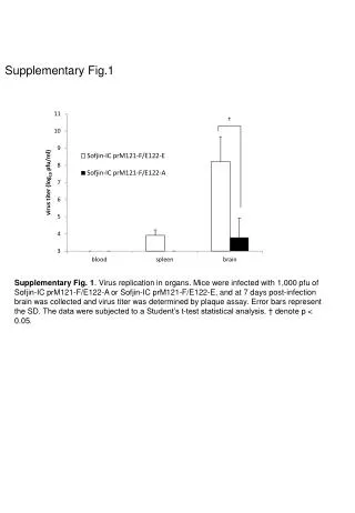
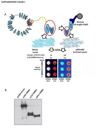
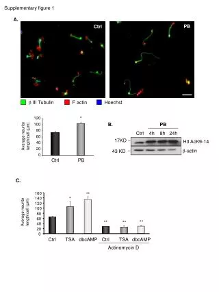
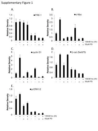
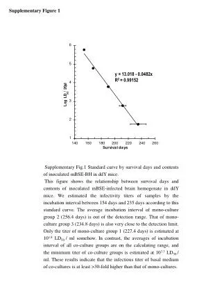
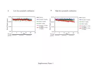
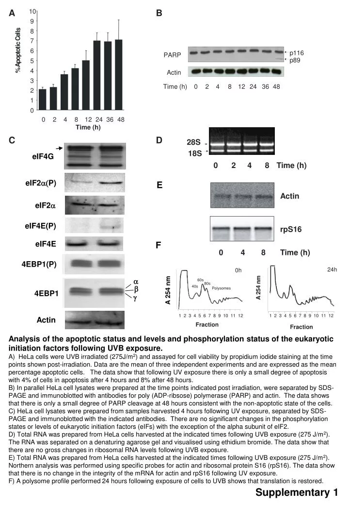
10 9 s 8 l l e C 7 p116 c PARP i t p89 o 6 t p o 5 p Actin A % 4 Time (h) 0 2 4 8 12 24 36 48 3 2 1 0 0 2 4 8 12 24 36 48 28S Time (h) 18S 0 2 4 8 Time (h) Actin rpS16 0 4 8 Time (h) 1 1 2 2 3 3 4 4 5 5 6 6 7 7 8 8 9 9 10 10 11 11 12 12 b g A B C D eIF4G eIF2a(P) E eIF2a eIF4E(P) eIF4E F 4EBP1(P) 24h 0h a m 60s n 80s 4 40s A 254nm Polysomes 4EBP1 5 2 A Actin Fraction Fraction Analysis of the apoptotic status and levels and phosphorylation status of the eukaryotic initiation factors following UVB exposure. A) HeLa cells were UVB irradiated (275J/m2) and assayed for cell viability by propidium iodide staining at the time points shown post-irradiation. Data are the mean of three independent experiments and are expressed as the mean percentage apoptotic cells. The data show that following UV exposure there is only a small degree of apoptosis with 4% of cells in apoptosis after 4 hours and 8% after 48 hours. B) In parallel HeLa cell lysates were prepared at the time points indicated post irradiation, were separated by SDS-PAGE and immunoblotted with antibodies for poly (ADP-ribsose) polymerase (PARP) and actin. The data shows that there is only a small degree of PARP cleavage at 48 hours consistent with the non-apoptotic state of the cells. C) HeLa cell lysates were prepared from samples harvested 4 hours following UV exposure, separated by SDS-PAGE and immunoblotted with the indicated antibodies. There are no significant changes in the phosphorylation states or levels of eukaryotic initiation factors (eIFs) with the exception of the alpha subunit of eIF2. D) Total RNA was prepared from HeLa cells harvested at the indicated times following UVB exposure (275 J/m2). The RNA was separated on a denaturing agarose gel and visualised using ethidium bromide. The data show that there are no gross changes in ribosomal RNA levels following UVB exposure. E) Total RNA was prepared from HeLa cells harvested at the indicated times following UVB exposure (275 J/m2). Northern analysis was performed using specific probes for actin and ribosomal protein S16 (rpS16). The data show that there is no change in the integrity of the mRNA for actin and rpS16 following UV exposure. F) A polysome profile performed 24 hours following exposure of cells to UVB shows that translation is restored. Supplementary 1