Download
1 / 4

E N D
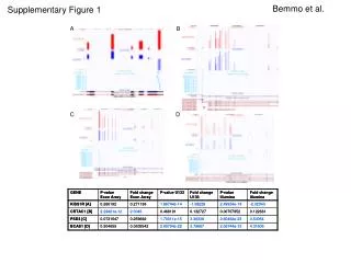
SUPPLEMENTARY FIGURE 1.Extraction of UV-induced excision products with different cell lysis methods.HeLa cells were exposed to 10 J/m2 of UV-C and then harvested 1 h after irradiation. Cells were lysed using one of the following lysis methods: A, Hirt procedure; B,a mild NP-40 lysis buffer (10 mMHepes, pH 7.6, 60 mMKCl, 1 mM EDTA, 0.1 % NP-40, and 1 mM DTT); C, RIPA buffer (25 mMTris-HCl, pH 7.6, 150 mMNaCl, 1% NP-40, 15 sodium deoxycholate, 0.1% SDS); D, a mild Triton X-100 lysis buffer (50 mMTris, pH 8.0, 150 mMNaCl, and 0.1% Triton X-100). E, a strong Triton X-100 lysis buffer (20 mMTris, pH 7.5, 150 mMNaCl, 1mM EDTA, 1mM EGTA, and 1% Triton X-100). SUPPLEMENTARY FIGURE 2. Degradation of excised oligonucleotides containing CPDs or (6-4)PPs. Primary and degraded forms of excised oligonucleotides containing CPDs (A) or (6-4)PPs (B) were quantified and are presented as means ± standard deviation (SD). The maximum values were set to 100 and the other values are presented relative to that value. SUPPLEMENTARY FIGURE 3. Quantification of excised oligonucleotides in UV-irradiated cells.A, Excised oligonucleotides were quantified in A375 cells 1 hr following exposure to 10 J/m2 of UV (original data is shown in Figure 6). Only those points that fall within the linear range of detection using the biotinylation and chemiluminescence technique are shown here. The fmol of excised oligomers was determined using a defined amount of a an oligonucleotide standard. Data are expressed as both mol per cell (mol/cell) and molecules of excised oligonucleotides per cell (molecules/cell). B, By calibrating the time course data in Figure 5 with the 1 hr time point data in A (82,000 excised oligomers/cell), the relative excision product signals for all of the other time points were converted to the number of molecules of excised oligomers per cell. C, Measurements of CPD and (6-4)PP repair in A375 cells exposed to 10 J/m2 of UV were obtained from our previous immunoslot blot data (10) and used to determine the number of excised oligonucleotides that are released from the genome. We determined that the aneuploid A375 cell line has 15 pg of DNA per cell (approximately 14 billion bp of DNA). Because a dose of 10 J/m2 of UV generates 1 photoproduct per 10 kb of genomic DNA (20), this dose results in the generation of 1.4x10⁶ photoproducts in the genomic DNA of A375 cells, of which approximately 80% are CPDs and 20% are (6-4)PPs. Using the actual or extrapolated genomic DNA repair values in (10), we then converted the relative CPD and (6-4)PP repair data into the number of molecules of CPD- and (6-4)PP-containing oligonucleotides that are cumulatively released as a function of time. D, The number of excised oligonucleotides per cell that are released as function of time was plotted for both the immunoslot blot (C) and Hirt extraction/3’-labeling (B) methods. E, The percentage of excised oligonucleotides that are released from the genome (C) that can be detected by Hirt extraction and 3’-labeling (B)was plotted for each time point.
A Supplementary Figure 3 C B D E