Download
1 / 38
380 likes | 385 Views
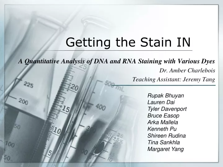
Getting the Stain IN. A Quantitative Analysis of DNA and RNA Staining with Various Dyes Dr. Amber Charlebois Teaching Assistant: Jeremy Tang. Rupak Bhuyan Lauren Dai Tyler Davenport Bruce Easop Arka Mallela Kenneth Pu Shireen Rudina Tina Sankhla Margaret Yang. Purpose.

E N D
Getting the Stain IN A Quantitative Analysis of DNA and RNA Staining with Various Dyes Dr. Amber Charlebois Teaching Assistant: Jeremy Tang Rupak Bhuyan Lauren Dai Tyler Davenport Bruce Easop Arka Mallela Kenneth Pu Shireen Rudina Tina Sankhla Margaret Yang
Purpose To identify dyesthat: • Can differentiate between DNA and RNAin gel electrophoresis • Are safe, inexpensive, and effective • Have a linear relationship between amount of nucleic acid and absorbance
Project Overview Nucleic Acid – Negatively Charged Polyacrylamide Gel Electrophoresis (PAGE) http://en.wikipedia.org/rna (2007). Retrieved July 29, 2008, from Southern Illinois University School of Medicine Web site: http://web.siumed.edu/~bbartholomew/images/chapter6/F06-21.jpg
(Begin Run) Loading Dye Loading Dye Loading Dye RNA DNA DNA RNA RNA Loading Dye RNA DNA
(destain in water) Staining the Gel DNA band 35 bases RNA band (18 bases)
Use of Fiber Optic Technology Halogen Source (2006, September 26). Introduction to the Ocean Optics Spectrometer System . Retrieved July 29, 2008, from Truman State University Chemistry Laboratory Web site: http://chemlab.truman.edu/Instrumentation/OceanOptics/OOIntro.htm Detector
Dyes Cresyl Violet Methylene Blue ToluidineBlue O Azure B Thionin Azure A Azure C Coomassie Brilliant Blue Brilliant Cresyl Blue Nile Blue A
DNA RNA General Testing of Dyes Strips of Gel Stained with Methylene Blue, Azure A, and Azure C prior to destaining
Azure B ––– RNA ––– DNA
Azure A ––– RNA ––– DNA
Azure C ––– RNA ––– DNA
Toluidine Blue O ––– RNA ––– DNA
Cresyl Violet ––– RNA ––– DNA
Brilliant Cresyl Blue ––– RNA ––– DNA
Thionin ––– RNA ––– DNA
Coomassie and Nile Blue A Coomassie Nile Blue A
Dyes Chosen for Quantitative Analysis Cresyl Violet Methylene Blue ToluidineBlue O Azure B Thionin Azure A Azure C Coomassie Brilliant Blue Brilliant Cresyl Blue Nile Blue A
DNA RNA Thionin 10 – 200 picomoles
DNA RNA Azure B 10 – 200 picomoles
Cresyl Violet DNA RNA 10 – 200 picomoles
DNA DNA RNA RNA Methylene Blue 10 – 200 picomoles
Conclusions • Methylene Blue: metachromatic, linear for RNA up to 100 picomoles • Azure B: shows promise for linearity for RNA • Thionin: abnormal pattern for DNA • Cresyl Violet: needs further research • All other dyes except Nile Blue and Coomassie show promise in nucleic acid staining
Future Research • More careful pipetting • The effect of dye band area on absorbance values • Mixing Dyes • Use of other statistical methods to determine linearity • Multi-linear regression pattern
Acknowledgements Dr. Miyamoto Dr. Paul Quinn Counselors Schering-Plough Foundation Novartis The Dorr Foundation The Jennifer A. Chalsty Foundation The Edward W. and Stella C. Van Houten Memorial Fund The Jewish Communal Fund Laura and John Overdeck NJGSS Alumni and Parents 1984-2008