Download
1 / 108
1.08k likes | 1.08k Views
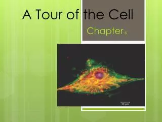
Explore the world of cells and their organelles through the use of microscopes. Discover the different types of microscopes and their capabilities, and learn about the process of cell fractionation. Understand the cell theory and the importance of cell size and diversity. Compare prokaryotic and eukaryotic cells and their structures. Dive into the fascinating world of cells with this comprehensive guide.

E N D
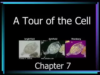
Microscopes • The discovery and early study of cells improved with the invention of microscopes in the 17th century. • In a light microscope (LM) visible lightpasses through the specimen and then through glass lenses. • The lenses refract light such that the image is magnified into the eye
Microscopes • Magnification is the ratio of an object’s image to its real size. • Resolving power is a measure of image clarity. • It is the minimum distance two points can be separated by and still be viewed as two separate points.
Microscopes • Light microscopes can magnify effectively to about 1,000 times the size of the actual specimen. • At higher magnifications, the image blurs
Microscopes • While a light microscope can resolve individual cells, it cannot resolve organelles. • To resolve smaller structures we use an electron microscope (EM), which focuses a beam of electrons through the specimen or onto its surface.
TEM • Transmission electron microscopes (TEMs) are used mainly to study the internal ultrastructure of cells. • A TEM aims an electron beam through a thin section of the specimen. • The image is focused and magnified by electromagnets. • To enhance contrast, the thin sections are stained with atoms of heavy metals.
SEM • Scanning electron microscopes (SEMs) are useful for studying surface structures. • The sample surface is covered with a thin film of gold. • The beam excites electrons on the surface. • These secondary electrons are collected and focused on a screen. • The SEM has great depth of field, resulting in an image that seems three-dimensional.
Electron Microscopes • Electron microscopes reveal organelles, but they can only be used on dead cells • Light microscopes do not have as high a resolution, but they can be used to study live cells. • Microscopes are a major tool in cytology, the study of cell structures. • Cytology coupled with biochemistry, the study of molecules and chemical processes in metabolism, developed into modern cell biology.
Isolating Cell Organelles • The goal of cell fractionation is to separate the major organelles of the cells so that their individual functions can be studied.
Cell Fractionation • This process is driven by an ultracentrifuge, a machine that can spin at up to 130,000 revolutions per minute • Fractionation begins with homogenization, gently disrupting the cell. • Then, the homogenate is spun in a centrifuge to separate heavier pieces into the pellet while lighter particles remain in the supernatant. • Repeating the process for longer & faster collects smaller organelles in the pellet
Cell Theory • Cells are the basic living units of organization and function • All cells come from other cells • Work of Schleiden, Schwann, and Virchow contributed to this theory • Each cell is a microcosm of life
Cell Size • Cell surface area-to-volume ratio • Plasma membrane must be large enough relative to cell volume to regulate passage of materials • Volume increases faster than surface area so cell must DIVIDE • Cell size and shape related to function
Prokaryotes & Eukaryotes • All cells are surrounded by a plasma membrane. • The semifluid substance within the membrane is the cytosol, containing the organelles. • All cells contain chromosomes which have genes in the form of DNA. • All cells also have ribosomes, tiny organelles that make proteins using the instructions contained in genes.
Prokaryotes & Eukaryotes • A major difference between prokaryotic and eukaryotic cells is the location of chromosomes. • In a eukaryotic cell, chromosomes are contained in a membrane-enclosed organelle, the nucleus. • In a prokaryotic cell, the DNA is concentrated in the nucleoid region without a membrane separating it from the rest of the cell.
Prokaryotes & Eukaryotes • In eukaryote cells, the chromosomes are contained within a membranous nuclear envelope. • The region between the nucleus and the plasma membrane is the cytoplasm. • All the material within the plasma membrane of a prokaryotic cell is cytoplasm.
Prokaryotes & Eukaryotes • Within the cytoplasm of a eukaryotic cell is a variety of membrane-bounded organelles of specialized form and function. • These membrane-bounded organelles are absent in prokaryotes.
Prokaryotes & Eukaryotes • Eukaryotic cells are bigger than prokaryotic cells • Ability to carry on metabolism set limits on cell size • Approximate Cell Size: • Smallest bacteria, mycoplasmas between 0.1 to 1.0 micron • Most bacteria are 1-10 microns in diameter, while Eukaryotic cells are typically 10-100 microns in diameter
The plasma membrane functions as a selective barrier that allows passage of oxygen, nutrients, and wastes for the whole volume of the cell.
Importance of Surface Area • The volume of cytoplasm determines the need for this exchange. • Rates of chemical exchange may be inadequate to maintain a cell with a very large cytoplasm. • The need for a surface sufficiently large to accommodate the volume explains the microscopic size of most cells. • Larger organisms do not generally have larger cells than smaller organisms - simply more cells.
Internal Membranes • A eukaryotic cell has extensive and elaborate internal membranes • Partition the cell into compartments • Many enzymes are built into membranes • Membrane compartments are involved in many METABOLIC functions
Membrane Structure • The general structure of a biological membrane is a double layer of phospholipids with other lipids and diverse proteins. • Each type of membrane has a unique combination of lipids and proteins for its specific functions. • For example, those in the membranes of mitochondria function in cellular respiration.
Nucleus • Contains most of the genes in a eukaryotic cell. • Some genes are located in mitochondria and chloroplasts. • Averages about 5 microns in diameter. • Separated from the cytoplasm by a double membrane. • These are separated by 20-40 nm. • Where the double membranes are fused, a pore allows large macromolecules and particles to pass through.
The nuclear side of the envelope is lined by the nuclear lamina, a network of intermediate filaments that maintain the shape of the nucleus.
Within the nucleus, the DNA and associated proteins are organized into fibrous material, chromatin. • In a normal cell they appear as a diffuse mass. • However when the cell prepares to divide, the chromatin fibers coil up to be seen as separate structures, chromosomes. • Each eukaryotic species has a characteristic number of chromosomes. • A typical human cell has 46 chromosomes, but sex cells (eggs and sperm) have only 23 chromosomes.
In the nucleus is a region called the nucleolus. • In the nucleolus, ribosomal RNA (rRNA) is synthesized and assembled with proteins from the cytoplasm to form ribosomal subunits. • The subunits pass from the nuclear pores to the cytoplasm where they combine to form ribosomes.
In the nucleus is a region called the nucleolus. • In the nucleolus, ribosomal RNA (rRNA) is synthesized and assembled with proteins from the cytoplasm to form ribosomal subunits. • The subunits pass from the nuclear pores to the cytoplasm where they combine to form ribosomes. • The nucleus directs protein synthesis by synthesizing messenger RNA (mRNA). • The mRNA travels to the cytoplasm and combines with ribosomes to translate its genetic message into the primary structure of a specific polypeptide.
Ribosomes • Ribosomes contain rRNA and protein. • A ribosome is composed of two subunits that combine to carry out protein synthesis.
Cell types that synthesize large quantities of proteins (e.g., pancreas) have large numbers of ribosomes and prominent nuclei. • Some ribosomes, free ribosomes, are suspended in the cytosol and synthesize proteins that function within the cytosol. • Other ribosomes, bound ribosomes, are attached to the outside of the endoplasmic reticulum. • These synthesize proteins that are either included into membranes or for export from the cell. • Ribosomes can shift between roles depending on the polypeptides they are synthesizing.
Many of the internal membranes in a eukaryotic cell are part of the endomembrane system. • These membranes are either in direct contact or connected via transfer of vesicles, sacs of membrane. • In spite of these links, these membranes have diverse functions and structures. • The endomembrane system includes the nuclear envelope, endoplasmic reticulum, Golgi apparatus, lysosomes, vacuoles, and the plasma membrane.
Endoplasmic Reticulum • The endoplasmic reticulum (ER) accounts for half the membranes in a eukaryotic cell. • The ER includes membranous tubules and internal, fluid-filled spaces, the cisternae. • The ER membrane is continuous with the nuclear envelope and the cisternal space of the ER is continuous with the space between the two membranes of the nuclear envelope.
There are two connected regions of ER that differ in structure and function. • Smooth ER looks smooth because it lacks ribosomes. • Rough ER looks rough because ribosomes (bound ribosomes) are attached to the outside, including the outside of the nuclear envelope.
The smooth ER is rich in enzymes and plays a role in a variety of metabolic processes. • Enzymes of smooth ER synthesize lipids, including oils, phospholipids, and steroids. • These includes the sex hormones of vertebrates and adrenal steroids. • The smooth ER also catalyzes a key step in the mobilization of glucose from stored glycogen in the liver.
Other enzymes in the smooth ER of the liver help detoxify drugs and poisons. • Also detoxifies alcohol and barbiturates. • Frequent exposure leads to the proliferation of smooth ER, increasing tolerance to the target and other drugs.
Rough ER is especially abundant in those cells that secrete proteins. • As a polypeptide is synthesized by the ribosome, it is threaded into the cisternal space through a pore in the ER membrane. • Many of these polypeptides are glycoproteins, polypeptides to which an oligosaccharide is attached.
Rough ER is especially abundant in those cells that secrete proteins. • As a polypeptide is synthesized by the ribosome, it is threaded into the cisternal space through a pore formed by a protein in the ER membrane. • Many of these polypeptides are glycoproteins, polypeptides to which an oligosaccharide is attached. • These secretory proteins are packaged in transport vesicles that carry them to their next stage.
Rough ER is also a membrane factory. • Membrane bound proteins are synthesized directly into the membrane. • Enzymes in the rough ER also synthesize phospholipids • As the ER membrane expands, parts can be transferred as transport vesicles to other components of the endomembrane system.
Golgi Apparatus • Many transport vesicles from the ER travel to the Golgi apparatus for modification of their contents. • The Golgi is a center of manufacturing, warehousing, sorting, and shipping. • The Golgi apparatus is especially extensive in cells specialized for secretion.
The Golgi apparatus consists of flattened membranous sacs – cisternae (looks like a stack of pita bread) • The membrane of each cisterna separates its internal space from the cytosol • One side of the Golgi, the cis side, receives material by fusing with vesicles, while the other side, the trans side, buds off vesicles that travel to other sites.