Download
1 / 56
560 likes | 629 Views
This week: Monday and Tuesday – Nervous System Overview ( Fucntions , Histology, Cell types) Wednesday and Thursday Nerve Firing Friday Assess – and Read . home Stretch!. Questions to answer by end of class. Name 2 divisions of nervous system

E N D
This week: Monday and Tuesday – Nervous System Overview (Fucntions, Histology, Cell types) Wednesday and Thursday Nerve Firing Friday Assess – and Read
Questions to answer by end of class • Name 2 divisions of nervous system • Name 2 divisions of peripheral nervous system • Name 2 divisions of Autonomic Nervous System • List functions of (formal) Nervous system
Today Nervous System Lecture 1 • Functions • Input, output, Interpret! • Organization • Where cells are found, and by what they do • Cells • Neurons • Supporting Cells • 6 new cells to ID
Functions of the Nervous System • Sensory input • Info gathered by sensory receptors • Integration • Interpretation of sensory input • Motor output • Activation of effector organs (muscles and glands) produces a response
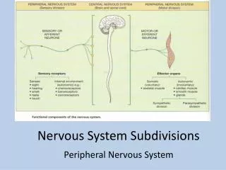
Sensory input Integration Motor output Figure 11.1
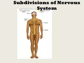
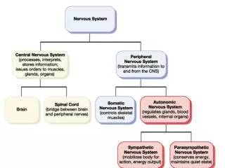
Divisions of the Nervous System • Central nervous system (CNS) • Brain and spinal cord • Integration & command center • Peripheral nervous system (PNS) • Paired spinal and cranial nerves carry messages TOand FROM the CNS
Peripheral Nervous System (PNS) • Two functional divisions • Sensory (afferent) division • Somatic afferent fibers—convey impulses from skin, skeletal muscles, and joints • Visceral afferent fibers—convey impulses from visceral organs • Motor (efferent) division • Transmits impulses from the CNS to effector organs
Efferent (Motor) Division of PNS • Somatic (voluntary Division)
Motor Division of PNS • Autonomic nervous system (ANS) • Visceral motor nerve fibers • Regulates smooth mm, cardiac mm, and glands • Stuff you are too busy to think about • Two functional subdivisions • Sympathetic • Parasympathetic involuntary division
There are 2 divisions of this Autonomic system • Sympathetic – fight or flight • Parasympathetic – rest and digest
Peripheral nervous system (PNS) Central nervous system (CNS) Cranial nerves and spinal nerves Brain and spinal cord Communication lines between the CNS and the rest of the body Integrative and control centers Sensory (afferent) division Motor (efferent) division Somatic and visceral sensory nerve fibers Motor nerve fibers Conducts impulses from the CNS to effectors (muscles and glands) Conducts impulses from receptors to the CNS Somatic sensory fiber Autonomic nervous system (ANS) Somatic nervous system Skin Visceral motor (involuntary) Somatic motor (voluntary) Conducts impulses from the CNS to cardiac muscles, smooth muscles, and glands Conducts impulses from the CNS to skeletal muscles Visceral sensory fiber Stomach Skeletal muscle Motor fiber of somatic nervous system Sympathetic division Parasympathetic division Mobilizes body systems during activity Conserves energy Promotes house- keeping functions during rest Sympathetic motor fiber of ANS Heart Structure Function Sensory (afferent) division of PNS Bladder Parasympathetic motor fiber of ANS Motor (efferent) division of PNS Figure 11.2
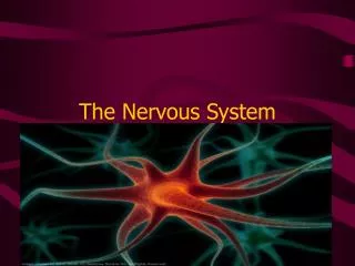
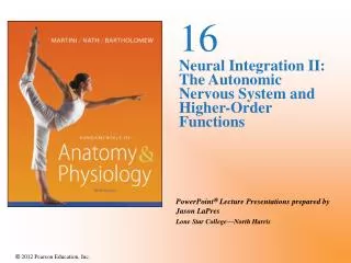
Histology of Nervous Tissue • Two principal cell types • Neurons—excitable cells that transmit electrical signals
Histology of Nervous Tissue • Neuroglia (glial cells)—supporting cells: • Astrocytes (CNS) • Microglia (CNS) • Ependymal cells (CNS) • Oligodendrocytes (CNS) • Satellite cells (PNS) • Schwann cells (PNS)
Astrocytes • Most abundant, versatile, and highly branched glial cells • Cling to neurons, synaptic endings, and capillaries • Support and brace neurons
Astrocytes • Help determine capillary permeability • Guide migration of young neurons • Control the chemical environment • Participate in information processing in the brain
Capillary Neuron Astrocyte (a) Astrocytes are the most abundantCNS neuroglia. Figure 11.3a
Microglia • Small, egg shaped cells with thorny processes • Migrate toward injured neurons • Phagocytize microorganisms and neuronal debris • Closest to immune cell you’ll get in CNS
Neuron Microglial cell (b) Microglial cells are defensive cells inthe CNS. Figure 11.3b
Ependymal Cells • Range in shape from squamous to columnar • May be ciliated • Line the central cavities of the brain and spinal column • Separate the CNS interstitial fluid from the cerebrospinal fluid in the cavities
Fluid-filled cavity Ependymal cells Brain or spinal cord tissue (c) Ependymal cells line cerebrospinalfluid-filled cavities. Figure 11.3c
Oligodendrocytes • Branched cells • Processes wrap CNS nerve fibers, forming insulating myelin sheaths
Myelin sheath Process of oligodendrocyte Nerve fibers (d) Oligodendrocytes have processes that formmyelin sheaths around CNS nerve fibers. Figure 11.3d
PNSSatellite Cells and Schwann Cells • Satellite cells • Surround neuron cell bodies in the PNS • Schwann cells (neurolemmocytes) • Surround peripheral nerve fibers and form myelin sheaths • Vital to regeneration of damaged peripheral nerve fibers
Satellite cells Cell body of neuron Schwann cells (forming myelin sheath) Nerve fiber (e) Satellite cells and Schwann cells (whichform myelin) surround neurons in the PNS. Figure 11.3e
Goals today: • Review organization of the Nervous system • Review Cell Types • Start Describing the neuron!
Neurons (Nerve Cells) • Special characteristics: • Long-lived ( 100 years or more) • Amitotic—with few exceptions • High metabolic rate—constant oxygen and glucose • Plasma membrane functions in: • Electrical signaling • Cell-to-cell interactions during development
Nerve parts • Pay attention to axon, dendrites, body
Cell Body (Perikaryon or Soma) • Its in the middle! Nucleus • Clusters of cell bodies are called nuclei in the CNS, ganglia in the PNS
Cell Body (Perikaryon or Soma) • Network of neurofibrils (neurofilaments) • Axon hillock—cone-shaped area from which axon arises
Dendrites (receptive regions) Cell body (biosynthetic center and receptive region) Nucleolus Axon (impulse generating and conducting region) Impulse direction Nucleus Node of Ranvier Nissl bodies Axon terminals (secretory region) Axon hillock Schwann cell (one inter- node) Neurilemma (b) Terminal branches Figure 11.4b
Processes • Dendrites and axons • Bundles of processes are called • Tracts in the CNS • Nerves in the PNS
Dendrites • “Cells little feelers” • Scattered branches • Receptive (input) region of a neuron • Convey electrical signals toward the cell body as graded potentials
The Axon • One axon per cell arises from the axon hillock • Long axons (nerve fibers) • Occasional branches ~90 degrees (axon collaterals)
The Axon • Numerous terminal branches (telodendria) • Knoblike axon terminals (synaptic knobs or boutons) • Secretory region of neuron • Release neurotransmitters to excite or inhibit other cells
Axons: Function • Conducting region of a neuron • Generates and transmits nerve impulses (action potentials) away from the cell body
Dendrites (receptive regions) Cell body (biosynthetic center and receptive region) Nucleolus Axon (impulse generating and conducting region) Impulse direction Nucleus Node of Ranvier Nissl bodies Axon terminals (secretory region) Axon hillock Schwann cell (one inter- node) Neurilemma (b) Terminal branches Figure 11.4b
Myelin Sheath • Lipid little blankets! • It functions to: • Protect and electrically insulate the axon • Increase speed of nerve impulse transmission
Myelin Sheaths in the PNS • Schwann cells wraps many times around the axon • Myelin sheath—concentric layers of Schwann cell membrane • Neurilemma—peripheral bulge of Schwann cell cytoplasm
Myelin Sheaths in the PNS • Nodes of Ranvier • Myelin sheath gaps between adjacent Schwann cells • Sites where axon collaterals can emerge
Schwann cell plasma membrane A Schwann cell envelopes an axon. 1 Schwann cell cytoplasm Axon Schwann cell nucleus 2 The Schwann cell then rotates around the axon, wrapping its plasma membrane loosely around it in successive layers. The Schwann cell cytoplasm is forced from between the membranes. The tight membrane wrappings surrounding the axon form the myelin sheath. Neurilemma 3 Myelin sheath (a) Myelination of a nervefiber (axon) Figure 11.5a
Unmyelinated Axons • Thin nerve fibers are unmyelinated • One Schwann cell may incompletely enclose 15 or more unmyelinated axons
Myelin Sheaths in the CNS • Formed by processes of oligodendrocytes, not the whole cells • Nodes of Ranvier are present • No neurilemma • Thinnest fibers are unmyelinated
Myelin sheath Process of oligodendrocyte Nerve fibers (d) Oligodendrocytes have processes that formmyelin sheaths around CNS nerve fibers. Figure 11.3d
White Matter and Gray Matter • White matter • Dense collections of myelinated fibers • Gray matter • Mostly neuron cell bodies and unmyelinated fibers
Structural Classification of Neurons • Three types: • Multipolar—1 axon and several dendrites • Most abundant • Motor neurons and interneurons • Bipolar—1 axon and 1 dendrite • Rare, e.g., retinal neurons