Download
1 / 6

E N D
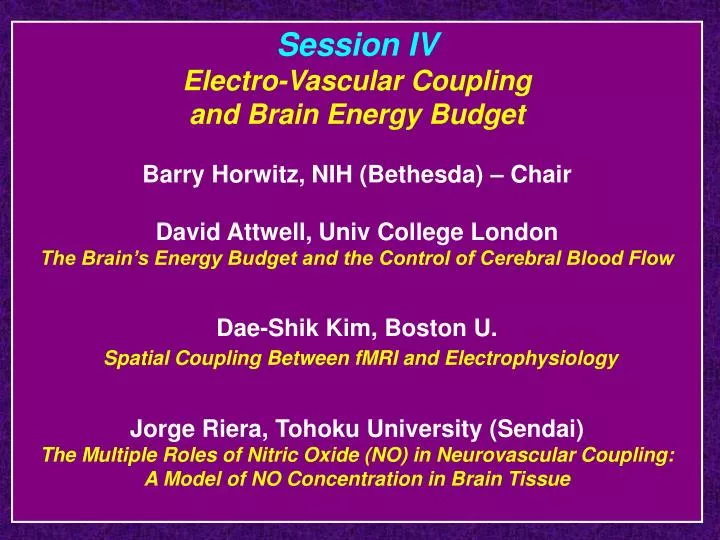
Session IVElectro-Vascular Coupling and Brain Energy BudgetBarry Horwitz, NIH (Bethesda) – ChairDavid Attwell, Univ College LondonThe Brain’s Energy Budget and the Control of Cerebral Blood FlowDae-Shik Kim, Boston U. Spatial Coupling Between fMRI and ElectrophysiologyJorge Riera, Tohoku University (Sendai)The Multiple Roles of Nitric Oxide (NO) in Neurovascular Coupling: A Model of NO Concentration in Brain Tissue
Why is this issue at a connectivity meeting? 1. Important for its own sake 2. Important because of its relation to interpreting fMRI and PET/rCBF data 3. Important because of its relation to neural modeling Recent efforts at large-scale modeling require integrating two models: 1. model of the neural activity 2. model that transforms the relevant neural activity to fMRI or PET signals (or EEG/MEG signals)
1993: Meeting on Neural Modeling and Functional Brain Imaging • Brought together modelers and functional brain imagers for the first time. • Tried to determine what research questions modelers could address • The four questions: • Relation between neural activity and imaging signals • Effective connectivity evaluation from imaging data • Integration of multimodality imaging data • Connecting multi-regional neural data to imaging data Horwitz, B. and Sporns, O.: Neural modeling and functional neuroimaging. Human Brain Mapping 1: 269-283, 1994.
Visual objects - Tagamets & Horwitz, Cerebral Cortex, 1998 Auditory objects - Husain et al., Neuroimage, 2004 Example Horizontal selective units Excitatory EE Inhibitory EI Horizontal selective units FS Corner selective units IT LGN D1 FR Vertical selective units Vertical selective units D2 Attention PFC V1/V2 V4 Model can do the following: 1. Simulate neural activity in each region 2. Simulate human behavioral performance 3. Simulate fMRI/PET activity in each region 4. Predicted existence of a type of neuron 5. Simulate MEG activity 6. Simulate functional & effective connectivity
Assumptions Used (1) BOLD and rCBF reflect the input to neurons; (2) excitatory and inhibitory inputs lead to increased BOLD and rCBF • fMRI • Integrate the absolute value of the synaptic activity over 50msec • Convolve with a hemodynamic response function (e.g., Boynton model) • Downsample every TR to get fMRI data • MEG • Local MEG signal is proportional to the difference between the excitatory and inhibitory synaptic activity on the excitatory units (e.g., pyramidal neurons) Some Issues Relevant to Modeling 1. What is the relation between neural activity and neuroimaging data? 2. Is it the same everywhere in the brain? 3. How do we take account of the activity in the different cortical layers? 4. There is a spread of neural activity due to local processing. Is it the same or different than the spread of hemodynamic change?