Download
1 / 42
560 likes | 1.44k Views
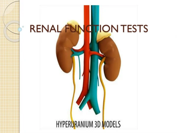
Biochemical Tests of Renal Function. D. Urinalysis Appearance Specific gravity and osmolality pH Glucose Protein Urinary sediments Measurement of GFR Plasma creatinine Clearance tests Tubular function tests. 2. Preanalytical phase of the urinary sediment. 2 nd morning void

E N D
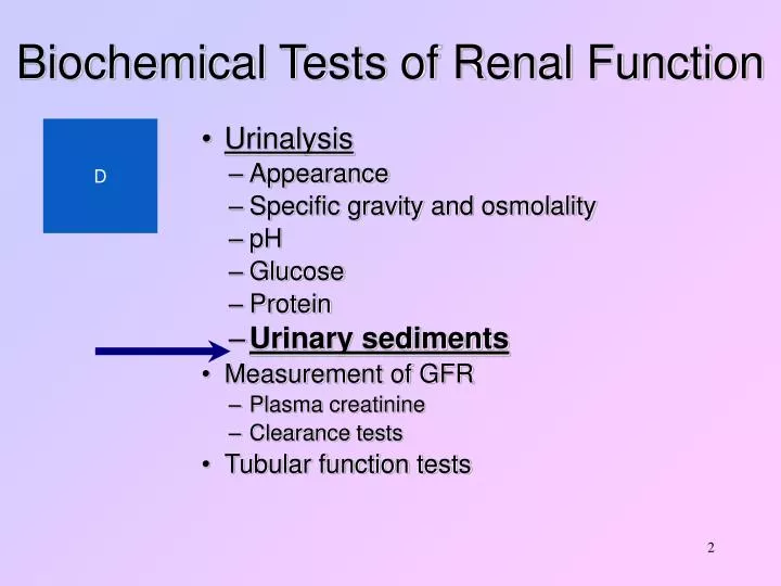
Biochemical Tests of Renal Function D • Urinalysis • Appearance • Specific gravity and osmolality • pH • Glucose • Protein • Urinary sediments • Measurement of GFR • Plasma creatinine • Clearance tests • Tubular function tests 2
Preanalytical phase of the urinary sediment • 2nd morning void • concentrated • acidic (pH>7 à lysis of WBC and casts) • without lysis of the elements (overnight) • Avoid physical exercise • Avoid excessive diuresis • No menstrual cycle • No catheterization 3
Stability of sample forthe urinary sediment • Collect in appropriate disposable containers • Analysis ideally within 1 hour (acceptable within 2 hour) to avoid bacterial overgrowth and lysis of cells and casts • Refrigeration and preservative agents are definitely inferior to prompt examination. 4
Preparation of theurinary sediment • Centrifugation • Supernatant removal • Resuspension • Preparation of slide • Evaluate immediately 5
Findings of the urinary sediment • Erythrocytes (morphology) • Leukocytes • tubular • Epithelial cells transitional • squamous • Casts • Crystals • Bacteria and yeasts 6
Urinalysis • Appearance: • Blood • Colour (haemoglobin, myoglobin) • Turbidity (infection, nephrotic syndrome) • Specific gravity • pH: • Physiological = acidic, except after meal 7
Urinalysis • At high density (>1025) RBC and WBC shrink • At low density (<1010) RBC and WBC swell and undergo lysis • At high pH (>7) WBC survival is shortened, casts diminishes and phosphates precipitate • At low pH (<5.5) urates precipitate 8
Microematuria • Microematuria: sangue nell’urina, in quantità ridotta, non visibile macroscopicamente • Rilievo: osservazione microscopica del sedimento e/o reazione positiva per l’emoglobina • Può essere presente per numerose cause fisiologiche (attività fisica!), ma deve essere sempre considerata con estrema attenzione • Alcuni dettagli morfologici delle emazie (emazie dismorfiche) possono consentire di identificare la sede di provenienza (d.d. glomerulo/basse vie urinarie) 9
Leukocytes (Neutrophils) • Urinary Tract Infections • Non-infectious renal diseases • glomerulonephritis • interstitial nephritis • polycystic kidney • tumours of urinary tract • urolithiasis • Contamination by genital secretions (women, + large amount of squamous cells) 13
Tubular cells • Acute tubular necrosis • Acute interstitial nephritis • Acute rejection of renal allograft • Active proliferative glomerulonephritis • Nephrotic syndrome • Nephrotoxic drugs 17
Transitional cells (of the deep layers) • Bladder carcinoma • Urolithiasis • Hydronephrosis • Uretheric stent • Bladder catheters 20
Tubular damage • Tubular cells • Casts (epithelial casts) • Dysmorphic erytrocytes • Lipids 21
Urothelium damage • Transitional cells of the deep • layers (ovoid and club-like) • Isomorphic erytrocytes • Leukocytes 22
Transitional cells (of the superficial layers) • Lower urinary tract infections 24
Squamous cells • Small number of SC are almost invariably present in the sediment of females, shed from the urethra and vagina • Large numbers of SC are seen in vaginitis often associated to bacteria and/or Candida. 26
Casts - I 27
Casts - II 28
Bacteria 40
Candidiasis 41
Urinalysis 3 • Urine sediments: • Microscopic examination of sediment from freshly passed urine: • Looking for cells, casts (Tamm-Horsfall protein), fat droplets • Red Cell casts - haematuria = glomerular disease • White Cell casts + polymorphs + bacteriuria = pyelonephrites • Lower UTI polymorphs no casts • Acute glomerulonephritis =haematuria, cells, casts • Chronic glomerulonephritis = less sediment 43