Download
1 / 36
360 likes | 930 Views
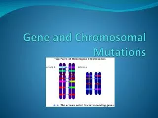
chromosomal mutations. Chromosomal mutations Changes in chromosome number Changes in chromosome structure Chromosome testing Karyotyping High resolution analysis Postnatal genetic testing. I. Chromosomal Mutations. possibly affecting more than one gene (multi-gene level)

E N D
chromosomal mutations • Chromosomal mutations • Changes in chromosome number • Changes in chromosome structure • Chromosome testing • Karyotyping • High resolution analysis • Postnatal genetic testing
I. Chromosomal Mutations • possibly affecting more than one gene (multi-gene level) • Changes in NUMBER • Monoploid number (2n – 1) • Euploidy (multiples of n) • Polyploid (3n, 4n, 5n…) • Triploid, tetraploid, pentaploid, hexaploid
1. Polyploidy • Usually lethalin mammals • Does occur in some animals - Reproduction via parthenogenesis, Flatworms, leeches, brine shrimp, lizards, salamanders, salmonids Polyploidy in plants:much more common because it can be tolerated by plants, can reproduce asexually… Important role in the evolution of plants – wheat: 2n = 14, 28, 42 chrysanthemum: 2n = 18, 36, 54, 72, 90
sympatric speciation: e.g. polyploidy in plants.. • Autopolyploidy: due to meiosis error. Offspring can self fertilize. • Allopolyploidy: • 2 different species mating, produce a hybrid that is polyploid: • The hybrid is fertile because the polyploid condition provides the homologous chromosomes for pairing during meiosis…
Endopolyploidy • only certain cells in the organism are polyploid • Liver cells, plant tissue (stem), larval gut tissue (mosquitos)
2. Aneuploidy – the total # is not an exact multiple of a set(2n +/- x) • Caused by Nondisjunction = failure of normal chromatid division during meiosis, two chromosomes go to one pole, none in the other. • Results in the wrong number of chromosomes. • Results in a gene imbalance
Fertilization of one of these affected gametes produces a zygote w/ either 3 members (trisomy) or only one member (monosomy) of the chromosome.
gene imbalance - THE problem Aneuploids are more abnormal than polyploids, why? (polyploid plants are completely viable and usually bigger, whereas in Drosophila the only aneuploids that survive are trisomics and monosomics for chromosome 4, the smallest chromosome) • Normal physiology of a cell depends on the proper ratio of gene products in the euploid cell. The amount of expression is correlated with the number of genes in a cell • If 3 copies present: 150% of the normal amount of protein will be made • If 1 copy present: 50% of the normal amount of protein will be made
Nondisjunction responsible for Turner’s syndrome and Kleinfelter’s syndrome… Turner’s syndrome produces sterile females with a normal # of autosomes and 1 X chromosome (XO). These are the only human monosomics that survive… Klienfelter’s syndrome individuals are trisomic: XXY, they are sterile males that are typically tall, and thin and some degree of mental retardation. XYY – trisomic males have mild mental retardation
Aneuploid Conditions in Humans Inherited disorders associated with aneuploidy. Trisomies and variations in the sex chromosomes result in mental retardation, organ defects, sexual immaturity, etc.
Trisomy 21, abnormal creases Trisomy 18, diaphragmatic hernia Turner’s syndrome, developmental abnormality polydactyly
Why is monosomy so bad? • Monosomics for all human autosomes die in utero • Any deleterious recessive alleles present on monosomic autosome will be automatically expressed
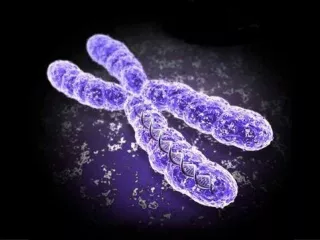
B. Changes in chromosome structure • A) Deletions • B) Duplications • C) Inversion • D) Translocation
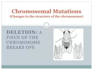
1. Deletions • Spontaneous breakage and rejoining • Interstitial deletion • Terminal deletion • Crossing over between repetitive DNA Region w/centromere usually maintained during division, the other part will be lost
Multigenic deletions • If both homologs have the same deletion then it will be lethal • If only on one homolog, the deletion can “uncover” lethal recessives in the heterozygous condition • Psuedodominance = when recessive alleles are expressed due to a deletion event
“partial monosomy” Caused by a heterozygous deletion of the tip of the p arm of chromosome #5 – phenotype: distinctive cat-like cry made by infants, microencephaly & moon-like face
2. Duplications • Extra copy of some particular region… Rare, and difficult to detect • Usually due to unequal crossing over during meiosis, or through replication error prior to meiosis • Not as problematic as deletions, but some problems are associated: • Bar eye in Drosophila (gene imbalance)
3. Inversions • Region breaks, rotates 180 degrees and rejoins • Generally viable, and show no abnormalities at the phenotypic level Paired homologs form an During synapsis, one chromosome must twist into a loop to pair up w/the genes on the other…
Types of inversions • 1) Paracentric – centromere outside of the inversion • Cross over products: dicentric and acentric chromosome • 2) Paricentric – inversion spans centromere • Cross over products: duplication, and deletion
During meiosis, homologs still pair up, even w/inversions -Inversion loop makes this possible
Crossing over produces affected chromatids: Duplication & Deletion events
4. Translocation-movement of chromosomal fragments to a new location. • Semisterility = an organism that is heterozygous for a reciprocal translocation usually produces about half as many offspring as normal • due to difficulty in chromosome segregation in meiosis. • Translocation cross = because of the translocations, the pairing of homologous regions leads to the unusual structure that contains four pairs of sister chromatids.
Nonreciprocal translocation – (unbalanced) • Centromeric regions of two nonhomologous acrocentric chromosomes become fused to form single centromere. -Down Syndrome chromosome 21 & 14 rearrangement leads to familial Down Syndrome. The heterozygote is normal, the 3 chromosomes must separate during meiosis (only 2/6 are normal, the rest either monosomic or trisomic) -Cancer (CML) type of leukemia, translocation between chromosome 9 & 22, leads to the movement of a gene where it will be overexpressed
Fragile sites – susceptible to breakage • Fragile X syndrome • Most common form of inherited mental retardation (1/4000 males, 1/8000 females) • FMR1 gene, has several trinucleotide repeats CGG in the 5’UTR region • Normal individuals = 6 to 54 repeats • Affected individuals = >230 repeats, region becomes modified (bases are highly methylated & gene NOT expressed) • Link between fragile sites & cancer • Chromosome #3 FRA3B region, FHIT gene often altered or missing in tumor cells taken from individuals w/ cancer
II. Chromosome Testing Chromosomes: • Karyotyping • High resolution chromosome analysis
A. karyotyping • adding a dye to metaphasic chromosomes; different dyes that affect different areas of the chromosomes are used for a range of identification purposes. • Giemsa dye is effective because it markedly stains the bands on a chromosome; Each chromosome can then be identified by its banding pattern • Amniocentesis • Chorionic Villi Biopsy
Prenatal genetic testing cont. Maternal Serum & Amniotic fluid • Alpha-fetoprotein (AFP) • Unconjugated estriol (uE3) • Dimeric inhibin A (DIA) • Fetal cell sorting
B. High resolution chromosome analysis • SKY – uses probes. Each of the individual probes complementary to a unique region of one chromosome - together, all of the probes make up a collection of DNA that is complementary to all of the chromosomes within the human genome. • Each probe is labeled with a fluorescent color that is designated for a specific chromosome.. • the probes hybridize, the fluorescent probes essentially paint the full set of chromosomes, can be analyzed to determine whether any of them exhibits translocations or other structural abnormalities.
2) In situ hybridization – used to map specific deletions & insertions No binding, 13.1-13.3 deleted
(FISH) analysis of a normal individual (D) and patient with a chromosome 22 deletion using a probe for the UFD1 gene. The patient has only one copy of UFD1 seen in blue (white arrows). Chromosome 22 was labeled with a red fluorescent marker (yellow arrows). http://www.ggc.org/clinical.htm