Download
1 / 58
580 likes | 592 Views
Structure determination by NMR. NMR principles Data acquisition Spectra process xwinnmr 、 nmrpipe 、 nmrview 、 Topspin Assignment Data Analysis Structure determination InsightII 、 Xplor 、 CNS Structural analysis Procheck 、 Molmol 、 Pymol. ~~NMR Experiments studies~~. Sample prepare.

E N D
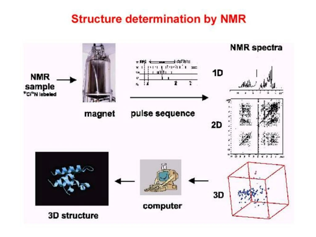
Structure determination by NMR • NMR principles • Data acquisition • Spectra process • xwinnmr、nmrpipe、nmrview、Topspin • Assignment • Data Analysis • Structure determination • InsightII、Xplor、CNS • Structural analysis • Procheck、Molmol、Pymol
Sample prepare • High concentrated protein • 10mg-30mg • Proton labeling • H1 • H1-N15 • H1-N15-C13 • Limitation • Protein molecular size <25 Kda
Modern Fourier transform NMR spectrometer Coil and superconductor LN2 and LHe2 tank
Spectra process and Assignment • Chemical shifts in proteins
Spectra process and Assignment • Chemical shifts in proteins the a-proton is always around 4 ppm; the aromatic protons are around 7 ppm; the backbone amides at 8 ppm.
Hα,H2O CH,CH2,CH3 HN, aromatic Well-dispersed 1D Spectrum
Why do we go beyond one dimension? • To resolve the crowded signals in 1D spectrum by spreading them into other dimensions. • To elucidate the “through-bond” and “through-space” relationships between the spins in the molecules.
COSY (correlation spectroscopy) • The original 2D experiment. Used to identify nuclei that share a scalar (J) coupling. The presence of off-diagonal peaks (cross-peaks) in the spectrum directly correlates the coupled partners. • NOESY (Nuclear Overhauser Effect Spectroscopy) • A 2D method used to map NOE correlations between protons within a molecule. The spectra have a layout similar to COSY but cross peaks now indicate NOEs between the correlated protons.
Secondary structure elements have characteristic NOE patterns
Spectra process and Assignment • 2D NMR spectroscopy
Spectra process and Assignment • 2D NMR spectroscopy • 2D TOCSY
Spectra process and Assignment • 2D NMR spectroscopy • 2D NOESY and TOCSY
Spectra process and Assignment • Assignment
Spectra process and Assignment • Assignment – TOCSY : identify spin system HN92 HN91 HN93 0 g b b b 4 0 a Ha91 a Ha93 Ha92 a 10 6 10 0 10 7
Spectra process and Assignment • Assignment - sequential assignment
Spectra process and Assignment • Assignment – NOESY : sequential assignment
Spectra process and Assignment • Assignment - sequential assignment
TOCSY : Amide to Aliphatic Region N’-ACGSC RKKCK GSGKC INGRC KCY-C’
H H H O N C C N C C N H O H NOESY and TOCSY : Amide to a Region
Isotope-labeling of proteins (I)15N labeling • Grow proteins on minimal media (M9) with 15NH4Cl as the sole nitrogen source. • $100-$1000 for mM sample. • Structure elucidation of medium-sized proteins (50-100 a.a.)
Isotope-labeling of proteins (II)15N, 13C labeling • Grow proteins on minimal media (M9) with 15NH4Cl as the sole nitrogen source and 13C-glucose as the sole carbon source. • $1000-$10000 for mM sample. • Structure elucidation of larger proteins (100-250 a.a.)
Isotope-labeling of proteins (III)15N, 13C, 2H labeling • Grow proteins on minimal media (M9) with 15N2H4Cl as the sole nitrogen source and 13C,2H-glucose as the sole carbon source in deuterated water. • Re-exchange deuterium on amide nitrogen to protons. • Strain must be adapted to grow on D2O. • > $10000 for mM sample. • Structure elucidation of larger proteins (> 200 a.a.)
Isotope-labeling of proteins (IV)Site-specific labeling • Add labeled amino acids to non-labeled media. • Assuming that the amino acid is not metabolized, all residues corresponding to that amino acid will be labeled in the protein. • Technique is interesting when structural or dynamic information is only required for specific residues. Thereby, the complete assignment of the protein may be circumvented.
Triple Resonance Experiment Use for Sequence Assignment • HNCA & HN(CO)CA • HNCO & HN(CA)CO • NHCBCA & CBCA(CO)NH
HNCOand HN(CA)CO Regions generated from Tyr56 to Glu63 are shown here. Red contours :former residues Black contours :intra-residues. Black and Red contours :intra- and inter-residue cross peaks are overlapping
HNCA and HN(CO)CA Regions generated from Tyr56 to Glu63 are shown here. Red contours :former residues Black contours :intra-residues. Black and Red contours :intra- and inter-residue cross peaks are overlapping
HNCACB + CBCA(CO)NH Regions generated from Tyr56 to Glu63 are shown here. The black lines show the scalar connectivities by Cα atoms and the blue lines show the scalar connectivities by Cβ atoms.
HCCH-TOCSY for side-chain proton assignments Strip plot extracted from a 3D HCCH-TOCSY spectrum obtained with uniformly 13C-labeled HP0495 in D2O
Structure determination by NMR • NMR principles • Data acquisition • Spectra process • xwinnmr、nmrpipe、nmrview、Topspin • Assignment • Data Analysis • Structure determination • InsightII、Xplor、CNS • Structural analysis • Procheck、Molmol、Pymol
Data Analysis and Structure determination • Data Analysis • NOESY – distance restrain • CSI – chemical shift index • Structure determination • principles
Data Analysis • NOESY
Data Analysis • NOESY medium range NOEs : i to i+2, i+3, i+4 long range NOEs : i to i+5……. filled circles < 6.0 Hz, open circles > 7.0 Hz filled diamonds kNH < 0.02 min-1
Data Analysis • NOESY H : Slowly exchanging (kNH < 0.02 min-1 ) amide protons the observed crosspeaks Hydrogen bonds NOE restrain : 20-30/per residues
Data Analysis • CSI
Data Analysis • CSI
Structure determination • Calculation • There is no method for a "direct" or ab initio calculation of a structure from NMR data. We have to include assumptions to make up the lack of experimental data. We therefore have to provide e.g. bond distances and angles for amino acids. • NMR structure calculation cannot result in the structure. Instead structure calculation is repeated many times, producing a large number of structural models. All the models that satisfy the experimental constraints are assumed as being representative of the protein.
Data Analysis • Calculation
Data Analysis • Calculation • Start: The temperature is set to 1000-3000 Kelvin which is very hot. At this extreme temperature different conformations of the polypeptide convert into each other very fast. In a completely random manner a large number of conformations are sampled. • We let the protein hop and shake around under these unnatural conditions to allow it to sample as many conformations as possible. The NOE distances are always switched on to force the protein to preferentially choose conformations that agree with the NOESY distances. • After a while the temperature is slowly reduced over quite some time to room temperature. While the system cools down we slowly reintroduce a correct description of the protein. • In the end, we simulate the protein as correct as it is possible on a computer. • The structure at the very end of the protocol is saved.