Download
1 / 11
110 likes | 220 Views
Deblurring in Atomic Force Microscopy (AFM) images. Supervisor: Prof. Anil Kokaram Co-Supervisor: Dr. David Corrigan Student: Yun feng Wang. Project Description. SFI project: collaboration with Nanoscale Function Group (NFG) in UCD

E N D
Deblurring in Atomic Force Microscopy (AFM) images Supervisor: Prof. Anil Kokaram Co-Supervisor: Dr. David Corrigan Student: Yun feng Wang
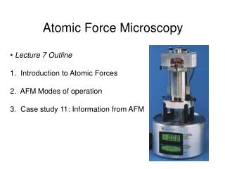
Project Description • SFI project: collaboration with NanoscaleFunction Group (NFG) in UCD • The principal measurement tool: Atomic Force Microscope (AFM) – artefacts • Project Target: AFM image restoration/artefacts removal
AFM Background • In this project, the image data was collected under liquid in 1mM HCL on a bespoke low noise AFM using nanosensor SSS-NCH probes.
The Subject – AFM imaging of amyloid fibrils • Approximately 20nm in diameter. In each example, slightly different copy of fibril with different brightness can be observed • Blurring artefact with a dramatic form distortion which is caused by the damage of the scanning probe • Use existing Bayesian deblurring algorithms in natural image domain to remove the blurring artefact in AFM images
An Initial Guess of Blur Kernel • As shown on right, a number of pixel pairs (highlighted in red) can be found with the same displacement (x, y) and intensity ratio µ between the fibril and its echo • Use Hough Transform technique to find a set of values (x,y, µ) which has the highest number of corresponding pixel pairs • The distortion in the imagespacewas modelled using the following equation: • where denotes the intensity of location (h, k) • is an echo of offset by a vector (x, y) • µ denotes the intensity ratio between the pixel intensity and its echo.
Hough Transform (HT) Results By applying Hough Transform, the image is then transform into a 3-D Hough Space. The bin with the highest number of votes denotes an initial guess of the blur kernel. A slice in the Hough Space containing the bin with the highest number of votes (dark red point). HT resultant kernel smoothed with a 7-tap Gaussian filter. Advantages: offers a good initial guess of blur kernel which can speed up the convergence & help with finding the global minimum instead of local minimum
Bayesian Deblurring • Nature Image Blur Model: • Blind Deconvolution: • Step1: Optimise latent image L with blur kernel k fixed • The latent image L can be optimised by finding the minimum of the function: • Solution: Fast TV-l1 Deconvolution method introduced in Xu 2010. • Step2: Optimise blur kernel k with latent image Lfixed • Novel blur kernel prior: assumes the new estimated blur kernel should be sparse and very similar to the HT blur kernel. • The blur kernel k can be optimised by finding the minimum of the function: • Solution: Rewritten as matrix multiplication form and optimised using the Conjugate Gradient (CG) based method introduced in Cho 2009. Latent Image Prior Likelihood/Noise Likelihood/Noise’s gradients Blur Kernel Prior
Limitations • The poorly deconvolved regions in the results (regions inside red window) are the regions in which the real fibril feature overlaps with its echoes • In this scenario, the AFM imaging process is thought to obey an overwrite model rather than a summation model of convolution as assumed in our algorithm
Conclusion & Future Work • As proved by the deblurring results, the proposed algorithm is successful at removing the large distortion artefact in AFM image. Also, the details inside the fibrils can be satisfactorily recovered with very few artefacts. • Adirection of future work is to investigate potential alternative ways of treating the overlap regions including investigating the possibility of a supervised deblurring algorithm