Download
1 / 83
870 likes | 1.22k Views
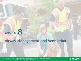
Chapter 9 Airway Management and Ventilation. Objectives (1 of 27). 1.6.11 Describe the anatomy of the following: upper airway, tongue, hypopharynx, nasopharynx, oropharynx, larynx, vocal cords. 1.6.12 Describe the function of the vocal cords.

E N D
Objectives (1 of 27) • 1.6.11 Describe the anatomy of the following: upper airway, tongue, hypopharynx, nasopharynx, oropharynx, larynx, vocal cords. • 1.6.12 Describe the function of the vocal cords. • 1.6.13 Describe the flow of air from outside the body into the trachea.
Objectives (2 of 27) • 1.6.14 Describe the reasons for and mechanisms of humidification and warming of the air as it passes through the naso- and oral pharynx. • 1.6.15 Describe the pathological conditions that can occur in the nose, pharynx, and larynx to obstruct or retard air flow and identify the complications of laryngeal fracture. • 1.6.16 Describe the methods of airway management.
Objectives (3 of 27) • 1.6.17 Describe the methods and management of an obstructed airway. • 1.6.18 Describe the mechanical methods of airway management including the benefits and limitations. Oral, nasal and EOA. • 1.6.19 Describe how the cervical spine is protected throughout these maneuvers.
Objectives (4 of 27) • 1.6.20 Describe the anatomy of the following: • Lungs • Trachea • Alveolus • Diaphragm • Thoracic wall • Pleural space • 1.6.21 Describe how pulmonary ventilation (inhalation and exhalation) is accomplished.
Objectives (5 of 27) • 1.6.22 Describe the gaseous exchange across the alveoli-capillary membrane (O2 and CO2.). • 1.6.23 Describe the pulmonary problems that can complicate exhalation and inhalation, the mechanisms by which they reduce ventilation and management of each problem, including: • Open pneumothorax • Diaphragmatic injury • Closed pneumothorax (simple and tension) • Flail chest.
Objectives (6 of 27) • 1.6.24 Describe the problems of ventilation. • 1.6.25 Define mouth-to-mask ventilation, its benefits and limitations. • 1.6.26 Discuss the bag-valve-mask (BVM), its benefits and limitations. • 1.6.27 Discuss the techniques for evaluating the effectiveness of ventilation. • 1.6.32 Discuss ventilation with an EOA.
Objectives (7 of 27) • *1.6.33 Discuss ventilation with an endotracheal tube. • *1.6.34 Describe the equipment and method of suctioning the airway, pharynx, and endotracheal tube. • S1.6.60 Demonstrate effective mouth-to-mask ventilation. • S1.6.61 Demonstrate effective bag-valve: • Mask • EOA • *ET
Objectives (8 of 27) • S1.6.63 Demonstrate the manual methods of airway management. • S1.6.64 Demonstrate the methods of management of an obstructed airway. • S1.6.65 Demonstrate the mechanical methods of airway management: • Nasal • Oral • EOA • *ET
Objectives (9 of 27) • S1.6.67 Demonstrate the use of various types of portable and fixed suction devices. • 1.7.1 Describe the anatomy of the following: upper airway, tongue, hypopharynx, nasopharynx, oropharynx, larynx, and vocal cords.
Objectives (10 of 27) • 1.7.2 Describe the relationship between: • Cords and larynx • Esophagus and larynx • Epiglottis and larynx • Tongue and larynx • True cords and false cords • Pharynx and larynx • 1.7.3 Given a list of arterial oxygen concentrations, the student should be able to select the normal PO2, for a young adult breathing air.
Objectives (11 of 27) • 1.7.4 Given a list of arterial carbon dioxide concentrations, the student should be able to select the normal PCO2. • 1.7.5 Given an increase in arterial PCO2, the student should be able to name this condition and describe its effect on respiratory activity and on blood pH in the normal individual.
Objectives (12 of 27) • 1.7.6 Given a decrease in arterial PO2, the student should be able to name this condition and describe its effect on respiratory activity in the normal individual. • 1.7.7 Given an increase in CO2 production, the student should be able to list at least two ways in which this increase may occur. • 1.7.8 Given an increase in CO2 elimination, the student should be able to describe how this elimination can occur.
Objectives (13 of 27) • 1.7.9 Given a list of statements, the student should be able to identify the statement that best describes the purpose of suctioning a patient. • 1.7.10 Given a diagram of a piston-powered suction unit, the student should be able to label and describe the operation and cleaning of each component and attached part.
Objectives (14 of 27) • 1.7.11 Given that there are various types of suction units, the student should be able to list at least four different types of units determined by the method in which the suction effect is obtained. • 1.7.12 Given that there are various types of suction catheters, the student should be able to list at least three different types, determined by difference in use and material composition.
Objectives (15 of 27) • 1.7.13 Given a list of situations describing patients who require suctioning, the student should indicate which type of catheter should be used. • 1.7.14 Given a list of statements, the student should be able to identify the statement that best describes the purpose of using the esophageal obturator airway.
Objectives (16 of 27) • 1.7.15 Given a list of situations describing patients with airway maintenance problems or potential airway maintenance problems, the student should be able to identify situations in which the use of the esophageal obturator airway is indicated and contraindicated. • 1.7.16 Given a list of situations, the student should be able to identify those situations in which the esophageal airway may be removed.
Objectives (17 of 27) • 1.7.17 Given a list of advantages, the student should be able to identify the advantages of using the esophageal obturator airway over other methods of airway control. • 1.7.18 Given a list of airway adjuncts, advantages, and disadvantages, the student should be able to match the airway adjuncts with the advantages and disadvantages.
Objectives (18 of 27) • S1.7.19 Given an adult manikin, oropharyngeal and nasopharyngeal airways, pocket mask, oxygen cylinder, and bag-valve-mask, the student should be able to demonstrate the procedure for administering intermittent positive pressure ventilation using: • Pocket mask • Bag-valve-mask and oropharyngeal airway • Bag-valve-mask with oxygen • Nasopharyngeal airway with bag-valve-mask
Objectives (19 of 27) • S1.7.20 Given a bag-valve-mask, the student should be able to demonstrate the assembly, disassembly, and cleaning of the bag-valve-mask unit. • S1.7.21 Given an adult manikin, an oropharyngeal airway, and a demand-valve unit, the student should be able to demonstrate the procedure for performing intermittent positive pressure ventilation.
Objectives (20 of 27) • S1.7.22 Given a demand-valve unit, the student should be able to demonstrate the assembly, disassembly, and cleaning of the unit. • 1.7.23 Given a list of disadvantages, the student should be able to identify the disadvantages of using the esophageal obturator airway over other methods of airway control.
Objectives (21 of 27) • 1.7.24 Given a diagram of the esophageal obturator airway, the student should be able to label and describe the function of all component parts. • 1.7.25 Given a list of equipment and materials, the student should be able to identify those items that must be available before esophageal obturation is begun.
Objectives (22 of 27) • 1.7.26 Given that a patient requires an esophageal obturator airway, the student should be able to list the procedures for insertion of the esophageal airway, including all steps in the proper sequence. • 1.7.27 Given a list of errors, the student should be able to identify common errors involved in the use of the esophageal obturator airway. • *1.7.28 Describe laryngoscope, suction, endotracheal tube, and bag-valve-mask.
Objectives (23 of 27) • *1.7.29 Discuss indications and contraindications of endotracheal intubation. • *1.7.30 Discuss alternatives to endotracheal intubation. • *1.7.31 Discuss skill deterioration and methods of prevention. • *1.7.32 Discuss need for rapid placement of the ET tube. • *1.7.33 Discuss methods of assuring and maintaining correct placement of the ET tube.
Objectives (24 of 27) • *1.7.34 Given that a patient needs suctioning and already has an endotracheal tube in place, the student should be able to describe the difference between endotracheal suctioning and oropharyngeal suctioning, including: • Dangers • Precautions
Objectives (25 of 27) • *S1.7.35 Given an adult intubation manikin, an esophageal obturator airway, 30cc syringe, and a bag-valve unit, the student should be able to demonstrate the technique for the insertion of an esophageal airway. He should further be able to demonstrate endotracheal intubation with the esophageal obturator in place and subsequent correct removal of the obturator. • *S1.7.36 Demonstrate placement of an ET within 45 seconds.
Objectives (26 of 27) • *S1.7.37 Demonstrate ventilation with a bag-valve and endotracheal tube. • *S1.7.38 Demonstrate method by assuring and maintaining correct placement of ET tube. • *S1.7.39 Demonstrate reventilation for missed intubation. • *S1.7.40 Demonstrate skills described above both on manikin and a live patient. • 1.8.18 Define acid-base balance.
Objectives (27 of 27) • 1.8.19 Discuss acid-base balance based on hydrogen concentration, pH, and buffer systems. • 1.8.20 Define and discuss the following: • Respiratory acidosis • Respiratory alkalosis • Metabolic acidosis • Metabolic alkalosis.
Airway • The most important steps in patient care are obtaining and maintaining a patent airway. • Oxygenation of the body’s tissues occurs through the processes of breathing and circulation. • The primary objective of emergency care is to ensure optimal ventilation. • Airway maneuvers are often neglected skills.
Anatomy of the Upper Airway (1 of 2) • Divided into upper and lower airways • Major functions of the upper airway • Warm, filter, and humidify air brought into the body • Pharynx • First portion of the upper airway • Composed of the nasopharynx and oropharynx
Anatomy of the Upper Airway (2 of 2) • Nasopharynx • Union of facial bones • Divided by septum • Contains the cilia and the turbinates • Oropharnyx • Oral cavity; begins with the mouth and teeth • Tongue, palate, epiglottis, and vallecula • Larynx • Complex structure • Glottic opening is the narrowest portion of the trachea
Anatomy of the Lower Airway • The function of the lower airway is to exchange oxygen and carbon dioxide. • Extends from C4 to the xiphoid process (externally); glottic opening to pulmonary capillary membrane (internally). • Inhalation • Surfactant • Atelectasis
Lung and Respiratory Volumes • Average adult male total lung capacity is 6 L • Tidal volume (average 5-7 mL/kg) • Dead space • Alveolar air • Minute volume • Functional reserve capacity • Residual volume • Inspiratory/expiratory reserve volumes
Ventilation (1 of 2) • Movement of air into and out of the lungs • Two phases of ventilation: • Inspiration • Stimulus to breathe comes from the respiratory center located in the medulla • Expiration • Stretch receptors in the chest wall and bronchioles send a signal to the apneustic center to inhibit the inspiratory center • Hering-Breuer reflex
Ventilation (2 of 2) • Respiration is the exchange of gases between a living organism and its environment. • Two types of respiration: • External – exchange of gases between the lungs and the blood cells • Internal – exchange of gases between the blood cells and tissues
Causes of Decreased Oxygen Concentration in the Blood • Any condition that reduces the surface area for gas exchange also decreases the oxygen supply • Pneumothorax • Decreased mechanical effort • Medical conditions • Pulmonary edema
Carbon Dioxide in the Blood • Fluctuates in relation to changes in breathing. • Hyperventilation rids the body of excessive amounts of carbon dioxide.
The Measurement of Gases • Dalton’s law of partial pressure • Total pressure of air (sea level) 760 torr • Major components • Nitrogen • Oxygen • Carbon dioxide • Water vapor
Respiratory Rate (1 of 2) • Neural control • Comes from medulla and pons • Apneustic center • Chemical stimuli • Chemoreceptors monitor the chemical composition of body fluids • Hypoxic drive is a “backup system”
Respiratory Rate (2 of 2) • Control of respiration by other factors • Increase or decrease respirations according to need. • Certain medications cause the respiratory rate to increase or decrease. • Respirations decrease as metabolism slows.
Airway Evaluation • Essential parameters • Bellows system • Normal rate: 12-20 breaths/min • Recognition of airway problems • Respiratory rate, regularity, or effort • Look, listen, and feel technique • History of present illness • Respiratory pattern changes
Acid-Base Balance (1 of 2) • Defining the acidity of a solution • Amount of free hydrogen ion • Ion shifts • Acid-base balance on both sides of the cell • Buffers • Imagined as a bucket • Circulating bicarbonate buffer • Holds and neutralizes excess acid
Acid-Base Balance (2 of 2) • Respiratory component • Fastest way to rid the body of excess hydrogen ions • Maintaining circulating levels of carbon dioxide in the blood • Renal component • Kidneys respond but take hours to days to restore pH • Excreting the acid
Compensatory Mechanisms • Acid-base disorders not immediately correctable by the body’s buffering systems initiate compensatory mechanisms to help return levels to normal.
Clinical Presentation (1 of 2) • Two types of acid-base disorders • Metabolic and respiratory • Respiratory acidosis • Always related to hypoventilation • COPD • Creates respiratory acidosis over time • Hypoxic drive stimulates breathing based on circulating oxygen levels in the bloodstream
Clinical Presentation (2 of 2) • Respiratory alkalosis • Always the result of hyperventilation • Metabolic acidosis • Any acidosis not related to the respiratory system • Metabolic alkalosis • Results from excessive loss of acid, either from urination or decreased acid levels in the stomach
Maintaining the Airway • Recovery position • Helps maintain a clear airway in a patient who has not had traumatic injuries and is breathing on their own with a normal rate and adequate tidal volume • Prevents aspiration of vomitus
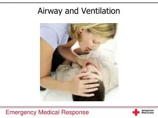
Airway Management • Emergency medical care begins with ensuring a patient’s airway is open and breathing is adequate. • The most common cause of airway obstruction in an unresponsive patient is the tongue.
Manual Maneuvers • Head tilt-chin lift maneuver • Jaw-thrust maneuver • Jaw-thrust with head tilt • Tongue-jaw lift maneuver