Download
1 / 37
470 likes | 747 Views
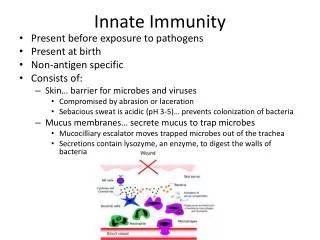
Inflammation and Innate Immunity (part I) . Inflammation Innate immunity and the initial response to infection Cytokines that induce inflammation and direct inflammatory cells Recognition of microbes by Toll-like receptors (TLRs) and other innate recognition elements

E N D
Inflammation and Innate Immunity (part I) • Inflammation • Innate immunity and the initial response to infection • Cytokines that induce inflammation and direct inflammatory cells • Recognition of microbes by Toll-like receptors (TLRs) and other innate recognition elements • Inflammation and recruitment of phagocytes • Uptake and killing of bacteria by phagocytes • Innate immunity against fungi, helminths, at mucosal epithelium
Inflammation Source: Wikipedia
Inflammation “rubor et tumor cum calore et dolore” (redness and swelling with heat and pain) --Cornelius Celsus in De Medicina, 1st century A.D. later “functio laesa” (disturbance of function) was added
Inflammation “rubor et tumor cum calore et dolore” (redness and swelling with heat and pain) --Cornelius Celsus in De Medicina, 1st century A.D. later “functio laesa” (disturbance of function) was added Inflammation is an adaptive response to noxious conditions (infection and tissue injury)--an attempt to restore homeostasis
Inflammation • Inflammation can be induced by immune recognition of infection or tissue damage (usually good) • Inflammation can be induced by immune recognition that is hypersensitive to environmental components or autoinflammatory or autoimmune (=disease)
Inflammation • Inflammation can be induced by immune recognition of infection or tissue damage (usually good) • Inflammation can be induced by immune recognition that is hypersensitive to environmental components or autoinflammatory or autoimmune (=disease) • Acute inflammation: influx of white blood cells and fluid from blood to fight infection and aid tissue repair • Chronic inflammation: inducer of inflammation is not removed • Leads to tissue damage and loss of tissue function (joint destruction, lung fibrosis, etc.) • Current view: aggressively fight inflammation in certain chronic diseases to decrease/delay progressive loss of function
Inflammation • Inflammation can be induced by immune recognition of infection or tissue damage (usually good) • Inflammation can be induced by immune recognition that is hypersensitive to environmental components or autoinflammatory or autoimmune (=disease) • Acute inflammation: influx of white blood cells and fluid from blood to fight infection and aid tissue repair • Chronic inflammation: inducer of inflammation is not removed • Leads to tissue damage and loss of tissue function (joint destruction, lung fibrosis, etc.) • Current view: aggressively fight inflammation in certain chronic diseases to decrease/delay progressive loss of function • Current research suggests that inflammation may play an important role in common chronic diseases including atherosclerosis, type 2 diabetes, neurodegeneration, and cancer
Immune sentinel cells in the tissues: dendritic cells Green= dendritic cells Blue= nuclei of all cells Langerhans cells (epidermal dendritic cells) in the skin WJ Mullholland et al. J. Invest. Dermatol. 126: 1541, 2006.
Infection leads to production of inducers of inflammation or dendritic cell Inflammatory mediators: Complex and many, but include: Lipids and Proteins (cytokines/chemokines) TNF Others
Cytokines • “Cytokines” are soluble protein mediators secreted by immune cells (mostly) that act on other cells to regulate their activity; many are called “interleukins” (IL-1, IL-2, etc.) (note: sometimes exist in cell-bound forms) • Cytokines have many functions, we’ll focus on a few central functions of some key cytokines (see “Cytokine primer” in syllabus/on iROCKET) • Name of a cytokine often doesn’t reflect its most important function (TNF stands for “tumor necrosis factor” but main function is to induce inflammation) • A subfamily of cytokines primarily functions in directing migration of cells, these are called “chemotactic cytokines” or “chemokines” Chemokines have systematic names: CCL1, 2, … and CXCL1, 2, … (but older names sometimes used, including IL-8)
The Initial Response to Infection: Innate Immunity • Recognition of infection by hard-wired recognition molecules or recognition of tissue damage and cell death, “danger” • Rapid mobilization of leukocytes to the site of infection and influx of plasma into the tissue site. (=inflammation) • Recruited innate immune cells kill microbes/virally infected cells (also can promote tissue repair but when dysregulated can exacerbate tissue injury) • Also, innate recognition promotes the adaptive immune response, which is slower but more powerful
Cytokines and Inflammation • Macrophages or DCs stimulated via innate immune receptors make pro-inflammatory cytokines, especially TNF (Tumor necrosis factor), IL-1, and IL-6 • TNF and IL-1 signal to endothelial cells to make them: • Leaky to fluid (influx of plasma; containing antibodies, complement components, etc.) • Sticky for leukocytes, leading to influx of first neutrophils, later monocytes, lymphocytes • IL-6 promotes adaptive immune responses and has systemic effects (“acute phase response” of liver, including C-reactive protein or CRP; levels used clinically as an indication of systemic inflammation)
Leukocyte recruitment to sites of inflammation or DC See Abbas and Lichtman Fig. 2-7 Note: molecular details of leukocyte extravasation will be covered in lecture Friday
Inflammation: Neutrophils vs. Monocytes • Acute inflammation is initially characterized as rich in neutrophils; later it is more monocytes and lymphocytes. This is controlled by which chemokines are expressed by the endothelial cells and by T cells. • Neutrophils are dedicated to killing microbes and are short-lived. They often damage host tissue as a byproduct. • Monocytes are multi-potential, depending on cytokine signals: +IFN-g: assume a vigorous killing phenotype similar to neutrophils +IL-4: “alternatively activated macrophages”; tissue repair, barrier immunity +IL-10: assume a wound-healing type phenotype (to clean up after infection is cleared) +GM-CSF: assume a dendritic cell phenotype and propagate adaptive immune responses
Anti-Inflammatory Therapeutics • NSAIDs: inhibitors of inflammation and fever (block prostaglandin synthesis) • Glucocorticoids are also potent anti-inflammatory drugs; natural systemic anti-inflammatory mechanism • Agents that block TNF are effective in treating rheumatoid arthritis, Crohn’s disease, etc. • Agents that block IL-1 are less effective for these diseases but are useful for some genetic inflammatory diseases (and are currently in clinical trials for more common conditions)
How is infection first recognized by the immune system? • What is seen by innate immunity? • Microbes evolve rapidly, so innate immunity must focus on highly conserved and essential components of microbes (cell wall structures; nucleic acids). So-called “Pathogen-associated molecular patterns” (PAMPs) • What mediates the recognition? • Diverse recognition elements; 4 key families of cellular receptors: • Toll-like receptors (TLRs; transmembrane receptors) • C-type lectin receptors (CLRs; transmembrane receptors) • RigI-like receptors (RLRs; cytoplasmic RNA helicases) • NOD-like receptors (NLRs; cytoplasmic sensors) • Also, recognition of molecules released from necrotic cells, tissue damage (“damage-associated molecular patterns” DAMPs or “danger”). Recognized by same families of innate receptors as PAMPs (Note:tissue damage can also be recognized by pain neurons, which can promote inflammation)
TLRs: Toll is required for innate defense in flies J. Hoffmann et al. Cell 1996
Toll-like receptors and recognition of pathogens ssRNA K. Takeda & S. Akira, Cell. Microbiol. 5: 143-53, 2003 Red circles: You should know these ligand/TLR pairs
Cellular localization of Toll-like receptors -Cell surface TLRs recognize bacterial cell wall structures-IntracellularTLRs recognize pathogen nucleic acids-Location likely aids discrimination of viral vs. host nucleic acids(likely connection with SLE)
The transcriptional activator NF-kB is a key player in inflammatory cytokine expression or TLR or IL-1 IKK= I-B kinase (Classical Pathway) Proteasome degrades I-B (Potential therapeutic target) Genes regulated: Inflammatory cytokines, chemokines, immune effector molecules, cell survival factors
Sepsis Syndrome: very bad(too much of a good thing) • Bacterial septicemia leads to activation of TLRs on monocytes in the blood, DCs in spleen • Systemic release of TNF and IL-1 leads to “inflammation” all over the body • Shock from loss of blood pressure (vasodilation and leakage of fluid into tissues) • TLRs also induce coagulation (via tissue factor) • Current therapy with some efficacy: “activated protein C”: promotes fibrinolysis, breaks down thrombi • The combination of effects frequently leads to multi-organ failure and death
Innate recognition by CLRs (examples) Geijtenbeek and Gringhuis, Nat. Rev. Immunol. 9: 465, 2009 NF-B
Innate recognition in the cytoplasm:NLRs and RLRs Modified from Abbas and Lichtman Fig. 2-2 Inflammatory cytokines Secrete anti-microbial peptides into lumen of crypts of sm. intest. (Mda5) Engulfment of bacteria invading the cytoplasm (autophagy) Interferon-
Common alleles of NOD2 are a genetic risk factor for Crohn’s disease • Several moderately common alleles of the NOD2 gene (7% of total alleles) increase susceptibility to Crohn’s disease (a form of inflammatory bowel disease) • Two copies of these alleles increase susceptibility by 40X • Mechanism: most evidence indicates these are loss-of-function alleles; unknown which function of NOD2 is most relevant
Processing of IL-1 and related cytokines: an important regulatory step • Some “NLRs” assemble to form the “inflammasome” which proteolytically processes IL-1 and related cytokines (IL-18) to their active, secreted forms. • Inflammasome in activated by cellular stress or recognition of microbial components in the cytoplasm
Processing of IL-1 and related cytokines: an important regulatory step • Some “NLRs” assemble to form the “inflammasome” which proteolytically processes IL-1 and related cytokines (IL-18) to their active, secreted forms. • Inflammasome in activated by cellular stress or recognition of microbial components in the cytoplasm • Genetic periodic fever syndromes are due to activating mutations in the inflammasome (active when it shouldn’t be) • Inflammasome is activated by some types of small crystals that can be phagocytosed by macrophages, important role in Gout
The NALP3-inflammasome activates caspase 1 in response to cellular insults Phagocytosed crystals Bacterial pore-forming toxins Efflux of K+ Bacterial flagellin Other insults/stresses Combinatorial Regulation of IL-1: • TLRs and NOD1/NOD2 induce synthesis of pro-IL-1 • Inflammasome processes it to generate active IL-1
Inducers of Inflammation • Ligands for TLRs, NOD1/2 or CLRs: DCs, macrophages makeTNF and IL-1 • Virus infections: infected cells, pDCs make IFN/ (type 1) • Tissue damage (cell necrosis etc.): “DAMPs” activate DCs, macrophages probably via TLRs, CLRs, inflammasomes (other receptors?) • Complement fragments (innate activators or IgM or IgG + antigen) • Mast cell activation (IgE+allergen or innate mechanisms: release histamine, leukotrienes, cytokines): eosinophil-rich inflammation (“type 2 immunity”) • Effector T cells responding to antigen (TNF + other cytokines; chemokines) -the 3 types of effector T cells induce inflammation of different characters (what white blood cells attracted)
Negative Regulation of Inflammation • Cells responding to innate stimuli stop making inflammatory mediators after short time period and convert to making anti-inflammatory lipids (resolvins, etc.) and anti-inflammatory cytokines (IL-10, TGF-) • But incoming inflammatory monocytes can still respond to stimuli if present and continue inflammation • Killing the infectious agent and removal of the dead cells, debris, crystals, etc. will stop stimulation of incoming inflammatory cells • Systemic elevation of inflammatory cytokines (esp. IL-1) induce production of glucocorticoids, which are anti-inflammatory (also by stress) • Regulatory T cells are also anti-inflammatory, both by blocking effector T cells and by inhibiting innate cells
Leukocyte recruitment to sites of inflammation: neutrophils are good at killing microbes or DC See Abbas and Lichtman Fig. 2-7
Phagocytosis and Killing of Microbes Abbas and Lichtman Fig. 2-9
Phagocytosis and Killing of Microbes Key Concepts related to phagocytosis: Opsonization: soluble immune recognition elements tag a particle for phagocytosis (opsonins include: IgG, C3b, Mannose-binding lectin, etc.) Interferon- from NK cell or Th1 cell promotes killing of internalized microbes by monocytes/macrophages Killing mechanisms: ROI, NO, proteases, anti-microbial peptides Abbas and Lichtman Fig. 2-9
Phagocytosis and Killing of Microbes Key Concepts related to phagocytosis: Opsonization: soluble immune recognition elements tag a particle for phagocytosis (opsonins include: IgG, C3b, Mannose-binding lectin, etc.) Interferon- from NK cell or Th1 cell promotes killing of internalized microbes by monocytes/macrophages Killing mechanisms: ROI, NO, proteases, anti-microbial peptides Genetic defects in phagocyte oxidase components: “chronic granulomatous disease” Abbas and Lichtman Fig. 2-9
Innate Immunity against fungal pathogens • CLRs are key innate recognition elements for fungi/yeast (TLRs can also play a role) • Neutrophils are important for killing most fungal pathogens • Some fungal pathogens can establish intracellular infections (like some bacterial pathogens): interferon- is important for defense; often also NO
Innate Immunity against helminths • Often multicellular parasites induce a “type 2” inflammation characterized by influx of eosinophils and basophils instead of neutrophils and monocytes • This type of inflammation is also seen in asthma and allergies, as will be discussed later in the course and can be propagated by Th2 adaptive immunity and/or IgE • Innate recognition is not yet understood, may include foreign polysaccharides (chitin), proteases, tissue damage • In some parasitic worm infections inside tissue, bacteria in the gut/feces of the worm stimulate TLRs and neutrophil-rich inflammation, which can cause pathology (African river blindness)
Innate Immunity and Mucosal Epithelium • Microbes are tolerated outside mucosal epithelium when consistent with its function (colon; upper airways) • Efforts to keep microbes out of some mucosal epithelial regions (small intestines and small airways) • Mechanisms include: actions of some surfactant proteins in lungs (bind to foreign polysaccharides); secretion of anti-microbial peptides by Paneth cells in crypts of small intestines; secretion of mucus by goblet cells; T cells in epithelial tissue; IgA • IL-13 is an important cytokine promoting mucus secretion